How was the feat of 'photographing men and women having sex with MRI' achieved?

by
The paper 'Men and women having sex were photographed by magnetic resonance imaging (MRI) ' was published in the British Medical Journal (BMJ) in 1999 and attracted worldwide attention. It summarizes how this research, which won the 2000 Ig Nobel Prize in Medicine, was carried out and published in a venerable medical journal.
Round the bend | The BMJ
https://www.bmj.com/content/367/bmj.l6654
This timeless piece of “body art” of people having sex in an MRI turns 20 | Ars Technica
https://arstechnica.com/science/2019/12/its-been-20-years-since-scientists-made-first-mri-images-of-human-copulation/
The BMJ is a year-end special issue published just before Christmas, and it is an annual event to publish several humorous papers. In the history of the Christmas issue that began in 1982, the most read by most people is a dissertation taken by MRI of men and women who have sex.
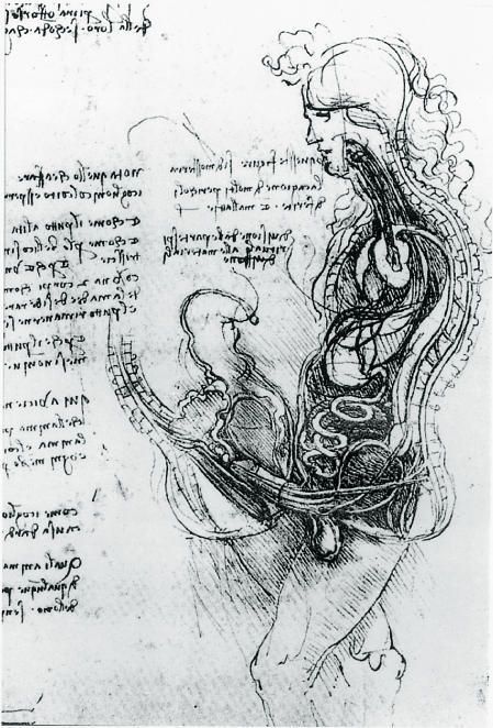
Sex is often viewed as taboo by conservatives, but in the olden days Leonardo da Vinci also wrote a note about men and women having sex. Da Vinci drew an anatomical cross-section of sexual intercourse in his 1493 memoir, 'The Copulation.' The memorandum on this sexual intercourse was not based on Da Vinci's own research, but was based on ancient Greek and Arabic texts. In addition, you can see from the image below that Da Vinci at this time draws the male genitals that entered the female vagina in a straight line.
In modern research, American obstetrician and gynecologist
Another well-known study of sex is the ' Masters Johnson Report ' by William Masters and Virginia Johnson. Masters and Johnson had hundreds of volunteers have sex in the lab and observed the condition.
And in 1999, Dutch physiologist Baek Van Andel published the world's first MRI scan of a man and a woman having sex. However, Van-Andel, who persuaded the hospital to get permission to use MRI, did not intend to take an image of sex from the beginning, but took 'an MRI image while a person is singing'. Was the first purpose. However, Van-Andel was inspired by the overly clear MRI image of the singer's throat that was actually shot, and came up with the project 'Sexing men and women with MRI.'

by Becca Tapert
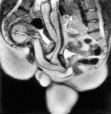
An anthropologist I knew, Ida Salives, was the first collaborative couple in Van Andel's research. In 1992, Mr. Salives and his girlfriend, Mr. Yap, went into a noisy MRI tube with a diameter of 50 cm and had sex together. Although he was worried whether he would really get an erection in the noisy MRI, Mr. Yap said he was able to get an erection safely.
This is the image published in the paper as a result of taking a picture with MRI for 45 minutes. You can see that Mr. Yap's penis on the right side is firmly inserted in Mr. Salives' vagina. From this image, Mr. Van Andel has been known so far that 'the penis at the time of insertion is not straight like the cross section drawn by Da Vinci, but actually bends like a boomeran'. I was able to discover a fact that was not there.
Van Andel, who thought the research had led to scientific results, sent the paper to the scientific journal Nature, but unfortunately it was rejected. Then, the Dutch tabloid squeezed the study and published it in paper, causing controversy such as 'How about using hospital MRI to shoot sex?', And the research team of Van Andel and others was in trouble. I will stand up.
Still, Van-Andel's enthusiastic persuasion managed to keep using the hospital's MRI, and by 1999 he continued his research with a total of eight couples working together. Eventually, Van Andel's paper was approved by the BMJ and will be published in the Christmas issue. The published paper attracted a great deal of attention and surprised the BMJ editors.
Twenty years have passed since the publication, and it seems that the memory of the editorial staff at that time is hazy, but the deciding factor for publishing the paper was that 'the composition of the cross section of Da Vinci and the MRI image was perfect. The editorial staff at that time recalled. There was also an opinion that images using new technology might be of interest to readers, and it was finally decided that 'Christmas issues that traditionally publish humorous papers can be published'. that's right.
Related Posts:


in Science, Posted by log1h_ik