'Nikon's Small World' 2020 award-winning works that compete for the beauty of micro and science
The results of ' Nikon's Small World ' 2020, which competes for images of the microscopic world that cannot be seen with the naked eye through a microscope, have been announced. The award-winning works that have been judged not only for the beauty of the images but also for their 'significance in science' are open to the public.
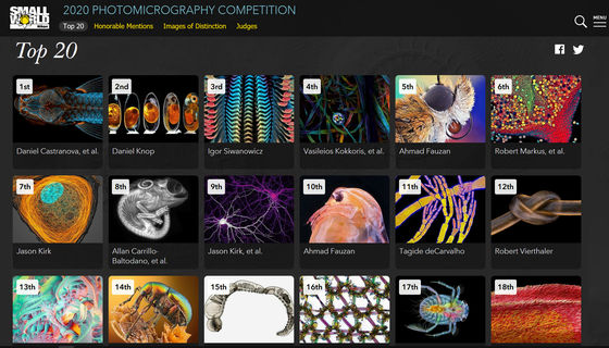
2020 Photomicrography Competition | Nikon's Small World
Glowing brains, fish embryos and a snail tongue taste success in microscopy photo contest | Live Science
https://www.livescience.com/nikon-small-world-photo-contest-winners.html
◆ 1st place: The back of the zebrafish juvenile
The first is a photograph of a zebrafish juvenile by Daniel Castlanova, Brandt M. Weinstein, and Baccary Samasa of the National Institutes of Health. It is created by stitching together more than 350 images taken by a confocal microscope that can take fluorescent images, and the skeleton is colored blue and the lymphatic system is colored orange.
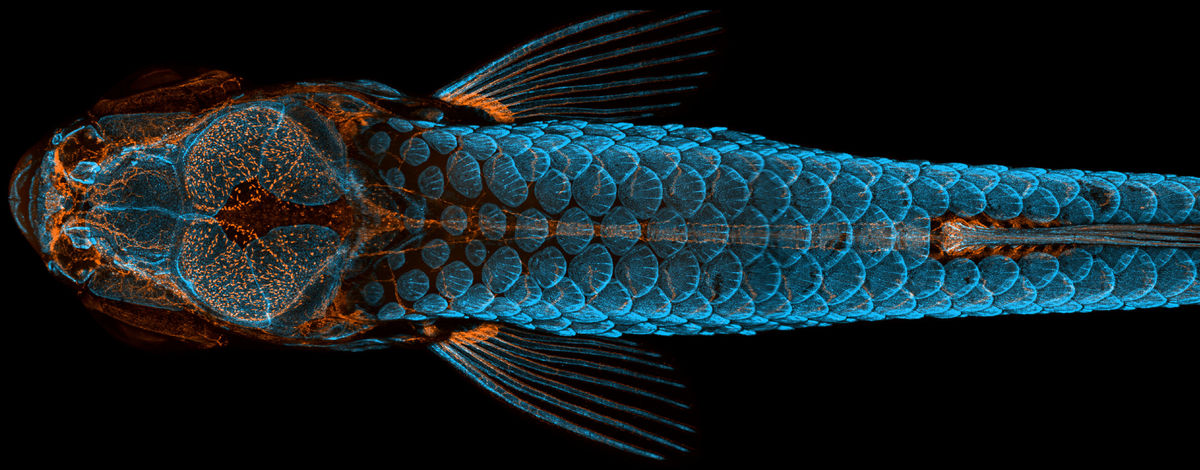
Regarding the images taken, Kastranova said, 'Not only are the images beautiful, but they also show how powerful the zebrafish lymphatic vessel development model is. This type of lymphatic system is thought to occur only in mammals. By continuing to study the lymphatic system of zebrafish, the scientific community has the potential to drive innovation in a variety of research and clinical trials, from drug testing to cancer treatment, because fish are better than mammals. It's much easier to breed. '
Dylan Burnett, one of the judges at Nikon's Small World, said, 'It takes an interesting scientific perspective to push images to the top in the judging. Kastranova's images are instantly clear, beautiful and scientifically meaningful. It was the only image that caught the attention of the judges and all the judges agreed. '
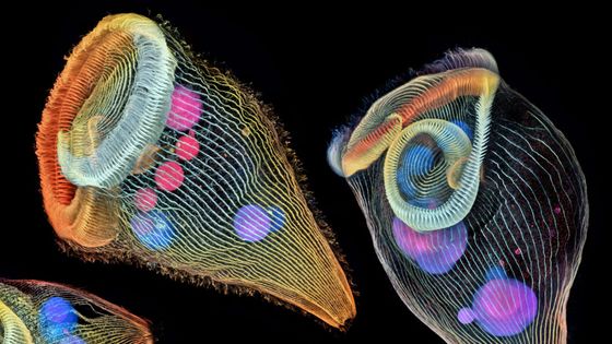
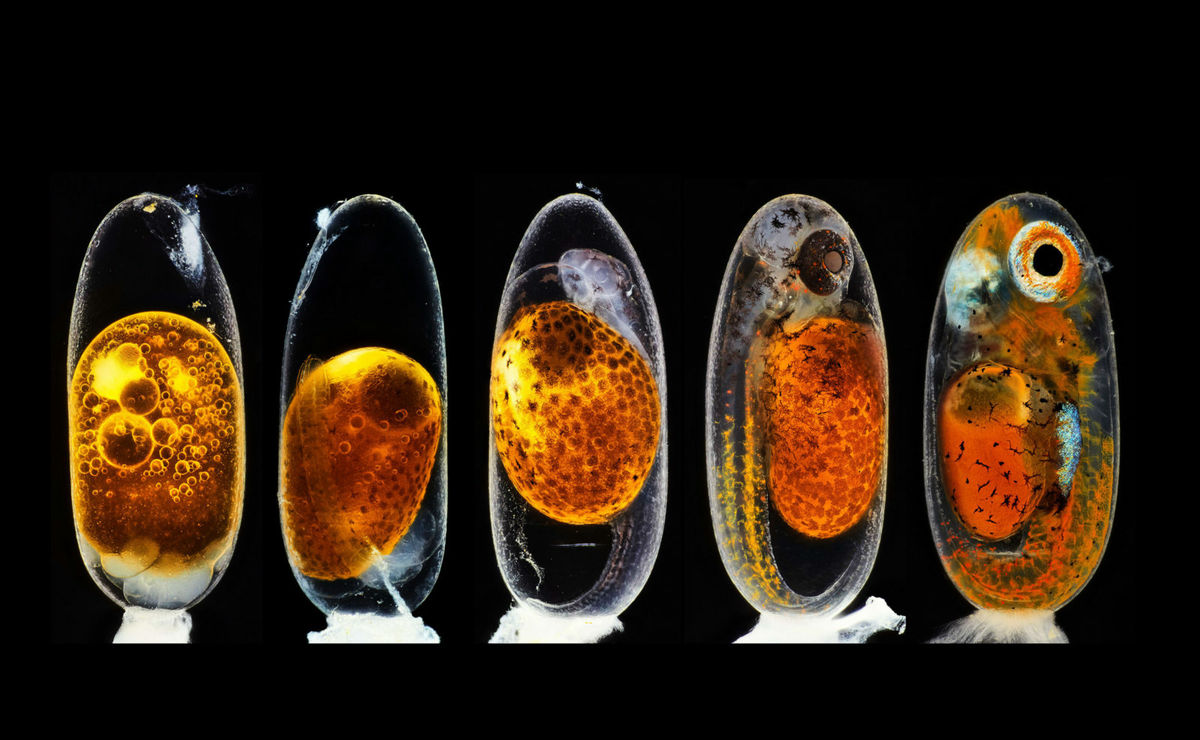
◆ 2nd place: Clownfish embryo
The following is a photograph of
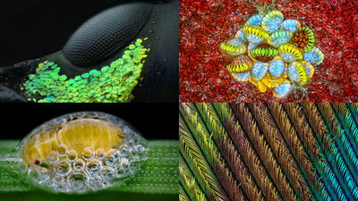
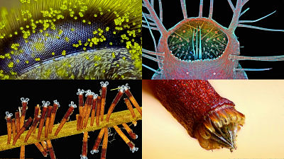
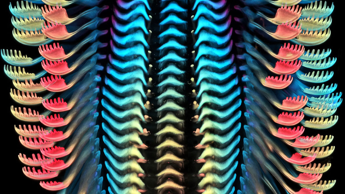
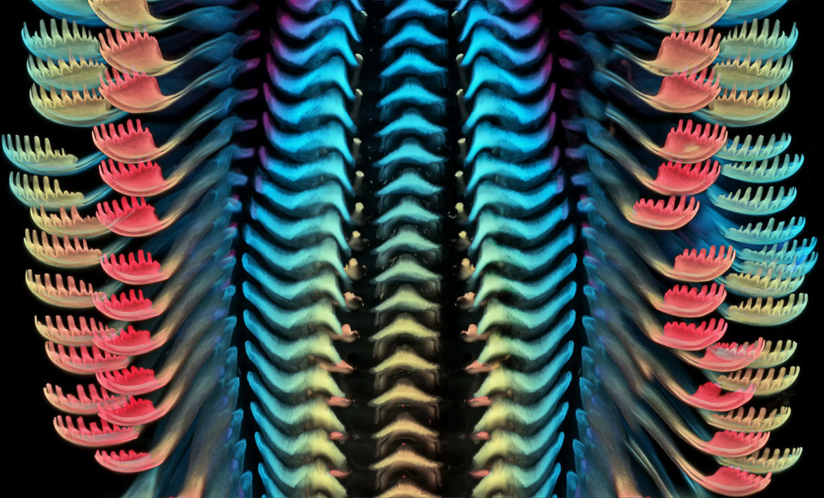
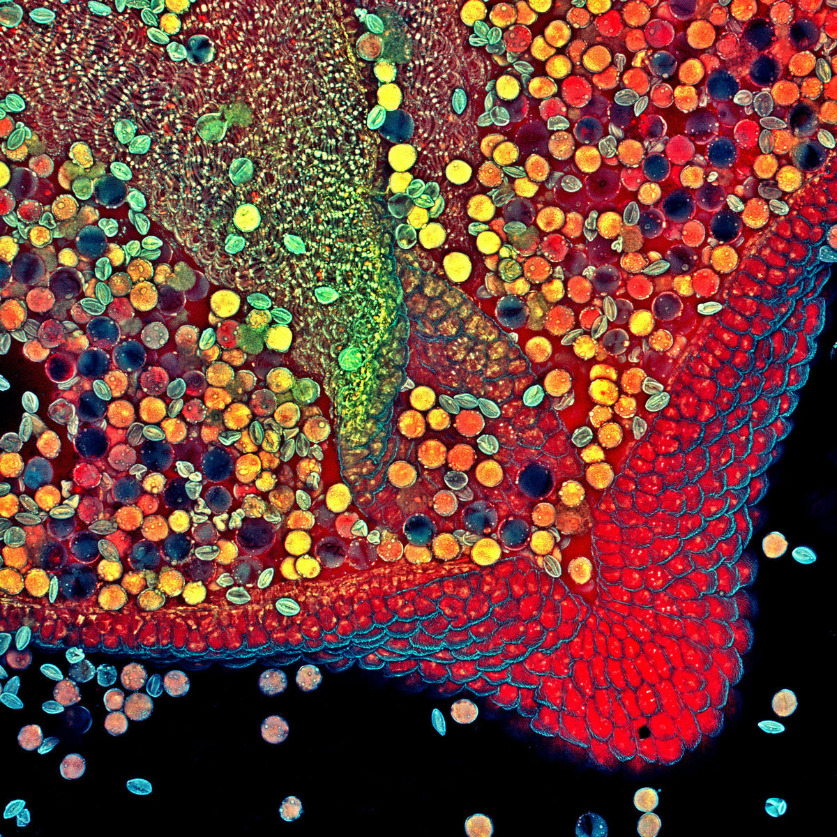
◆ 3rd place: Snail's Radula
It looks like a colorful feather, but it's actually a snail's
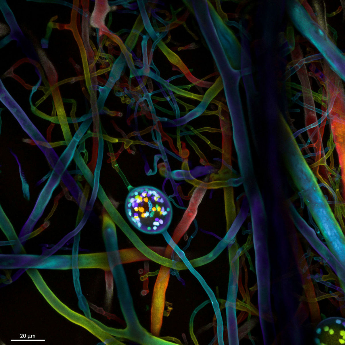
◆ 4th place: Arbuscular mycorrhizal fungus nuclei and hyphae
◆ 5th place: Bogong moth head
According to one theory, the Bogong moth , which is said to 'taste popcorn,' was shot at 5x magnification in the image below. The Bogong moth, photographed by Saipem's Ahmad Forzan, is captured in detail, including compound eyes and individual hairs.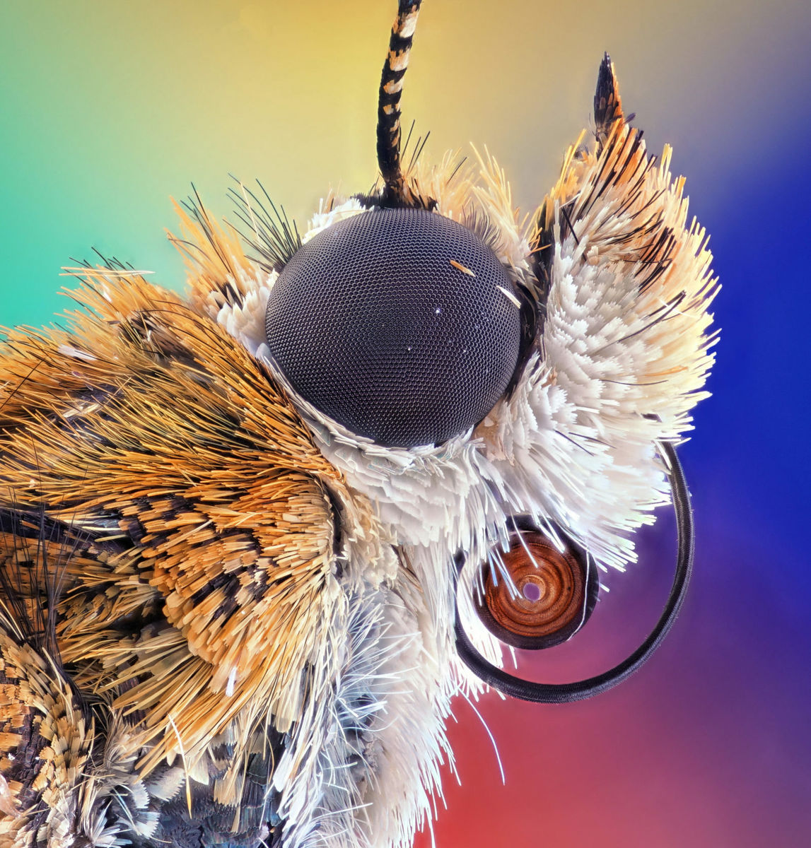
◆ 6th place: Hebe's fast
It is a mysterious image of colorful grains of red, blue, and yellow, but this is called '
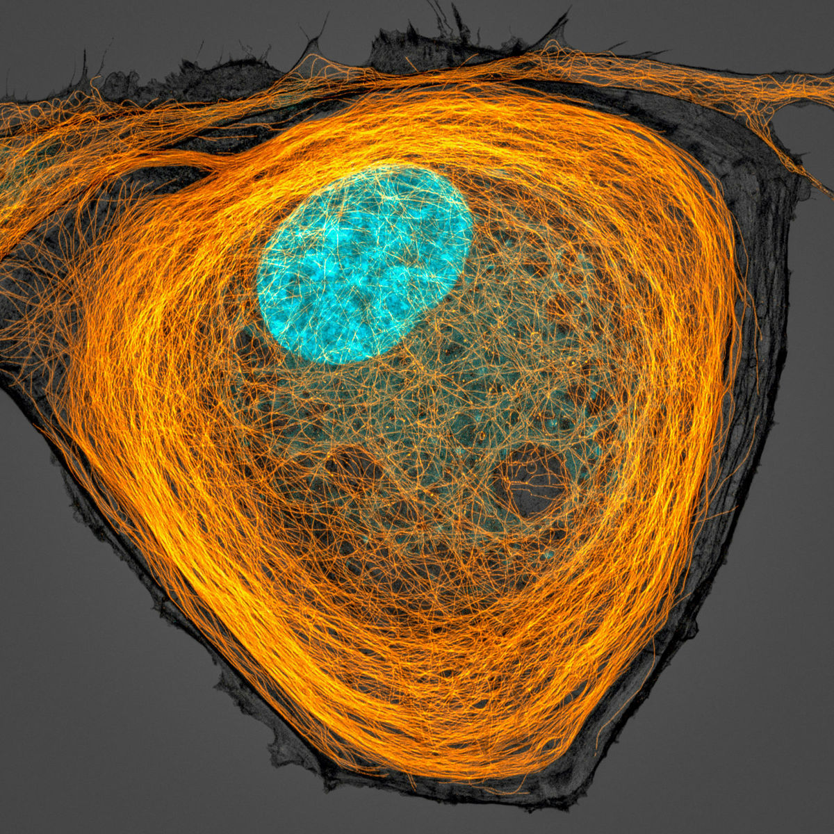
◆ 7th place: Intracellular microtubules
Below is a 63x magnified cell photographed by Jason Kirk of Baylor College of Medicine. The intricately intricate orange thin lines are the cell
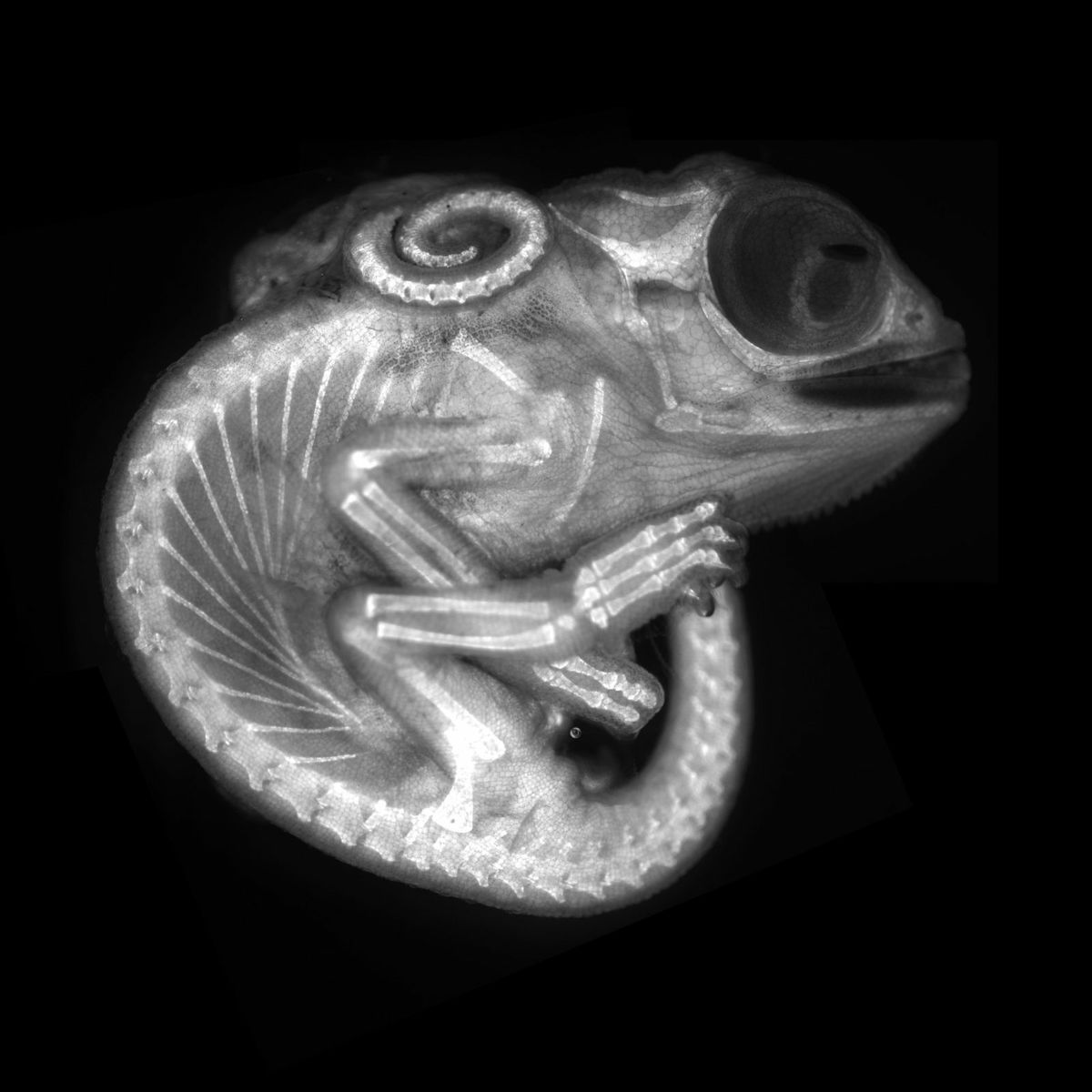
◆ 8th place: Chameleon embryo
The following image shows a chameleon embryo that has already grown to look like an adult chameleon at a magnification of 10x. Photographed by Alan Carilo and others from the Department of Biochemistry, Queen Mary University.
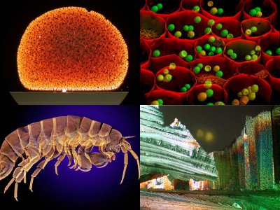
◆ 9th place: Neuron connection in the hippocampus
The white highlight in the image is a

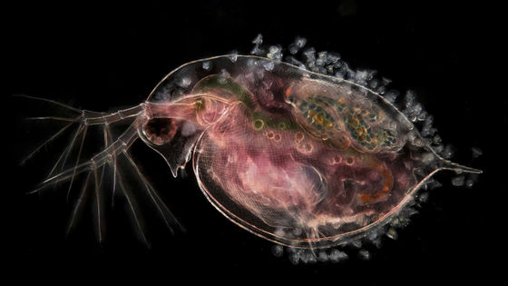
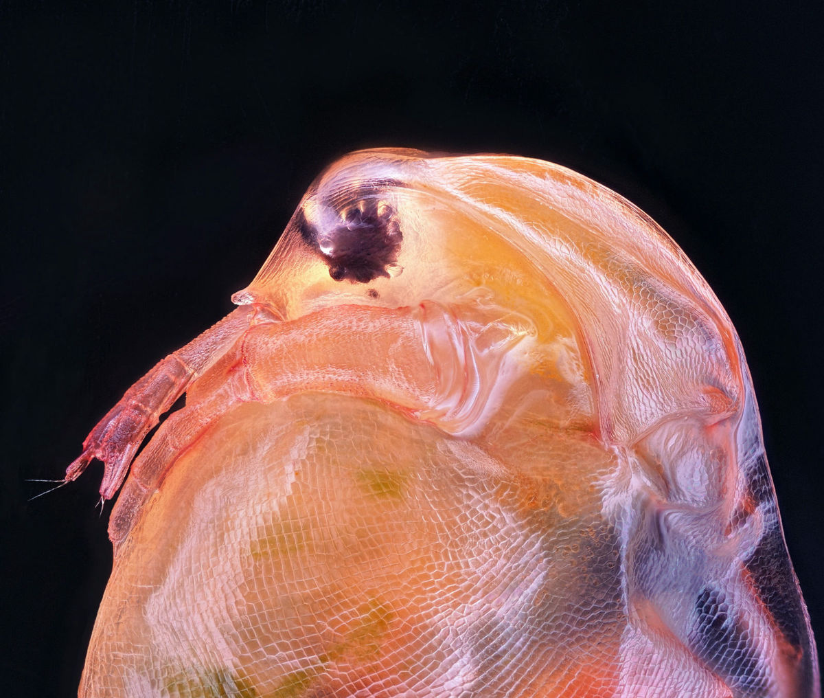
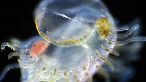
◆ 10th place: Daphnia magna
In the image below,
In addition to the works introduced in the article, the award-winning works and winning works after the 11th place are published on the official page below.
2020 Photomicrography Competition | Nikon's Small World
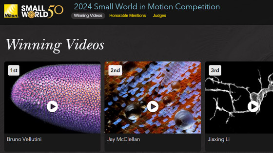
At 'Nikon's Small World' in 2020, a contest for the movie category was held in addition to the photo category. The results of the movie section can be seen in the following articles.
An excellent work of the contest 'Nikon's Small World In Motion 2020' that captures the fantastic micro world with a movie will be announced --GIGAZINE
Related Posts: