A structure hidden in the retina of the eye optimized the function of the eye
ByBen K Adams
The innermost retina of the eyeball is an organ that changes the visual image composed of light information into a neural signal and emits a signal to the brain and it is generally known that it functions like a camera film It is. The structure of the retina is sometimes optimized to maximize the work of the eyes, and Dr. Erez Ribak of the Israel Institute of Technology, who discovered that fact, explains by himself.
Look, your eyes are wired backwards: here's why
https://theconversation.com/look-your-eyes-are-wired-backwards-heres-why-38319
Phys. Rev. Lett. 104, 158102 (2010) - Retinal Glial Cells Enhance Human Vision Acuity
http://journals.aps.org/prl/abstract/10.1103/PhysRevLett.104.158102
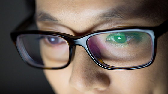
The retina in the eyeball is divided into several layers, and behind the retina reacts to the colors red, green and bluePyramidal photoreceptorAlthough it can not distinguish colors, it is highly sensitive to light and functions in the darkRod photoreceptorthere is. Conical photoreceptor cells and rods There are several layers consisting of cells such as ganglion cells and amacrine cells on the near side, that is, on the cornea side, and light entering from the eye passes through several layers It is to arrive at cone photoreceptor cells and rod photoreceptors, but why there is a layer composed of cells on the front side rather than behind the cone photoreceptor cells and rod photoreceptors is important for researchers But he seems to have been considered a mystery for many years.
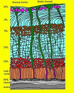
In order to unravel the mystery related to the structure of the retina, Dr. Ribak of the Israel Institute of Technology made a model of the retina in 2010 and carried out experiments, and the glial cells with the highest density among the cells present in the retina display the clarity of the vision We found that there is an increasing effect. However, what I was really interesting in experimenting is that the green and red light were more intensive than the blue and transmitted through the glial cells. Also in simulation experiments using computers, it turned out that green and red passing through glial cells was 5 to 10 times more concentrated, that is, when blue passes.
Next, Dr. Ribak conducted an experiment using guinea pigs to investigate how light is transmitted through the retina. The experiment is to apply light of 27 colors to the retina of the guinea pig and to examine the state with a microscope. Experimental results showed that not all light was evenly dispersed when passing through the retina but concentrated in a specific area. The part where the light was concentrated is thin and has a vertically elongated shape and extends from the entrance of the retina from the entrance of the retina to the pyramidal photoreceptor cell and the rod photoreceptor cell, and the concentration of light passing therethrough is the highest, about 10 normal It is said that it doubled.
ByThomas Tolkien
It was also found that glial cells play the role of guiding red and green light to optimal pyramidal photoreceptors, respectively. On the contrary, the blue light was dispersed and reached the rod photoreceptor without glial cells functioning well.
Simply summarizing the experimental results, "The layers in front of the cone photoreceptor cells and rod photoreceptors are responsible for guiding red and green light to the cone visual cells and rod photoreceptors more concentrated than blue It is said that it is fulfilled. " Especially red and green are colors which are recognized many in the daytime when the sun is rising and are colors which are not needed much in the evening. Compared with daytime and nighttime, daytime people use eyeball frequently, so it can be said that the retina is optimized so that the eyeball can distinguish red and green more.
Related Posts:


in Science, Posted by darkhorse_log