Successfully visualize how the mouse embryo changes into an organ
From the discovery of ES cells and IPS cells, studies of making organs from cells have become active. Meanwhile, a technique called " Adaptive light-sheet microscopy " appeared that images the appearance of cells changing from embryo to organ after fertilization.
In Toto Imaging and Reconstruction of Post-Implantation Mouse Development at the Single-Cell Level: Cell
https://www.cell.com/cell/fulltext/S0092-8674(18)31243-1
New Microscope Offers 4-D Look at Embryonic Development in Living Mice | Janelia Research Campus
https://www.janelia.org/news/new-microscope-offers-4-d-look-at-embryonic-development-in-living-mice
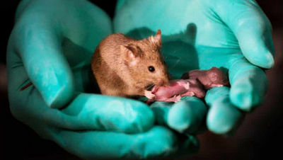
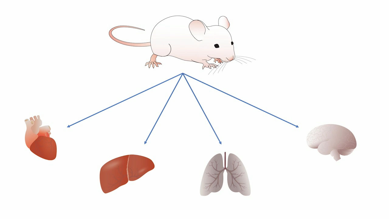
Understanding how early cells differentiate after fertilization is important because it is expected to be applied to regenerative medicine etc. which produces specific organs using stem cells. Therefore, techniques to visualize how cells differentiate have been studied for a long time. Dr. Janela Keller's team at Howard Hughes Medical Institute had already succeeded in photographing zebrafish in 2008 and flies in 2014 about embryo differentiation, respectively. However, while the embryos of these organisms were simple and easy to observe from the outside, it was difficult to photograph mammalian cell differentiation.
In the case of mouse embryos, it is not only difficult to grow embryos under the conditions of the laboratory without death, but also the initial organ formation begins 48 hours after fertilization, the rate of growth is higher than that of flies It was fast, so it was difficult to shoot the state of growth. Also, as the embryo continually changes its movement position with growth, it was impossible to keep focusing on the embryo manually.
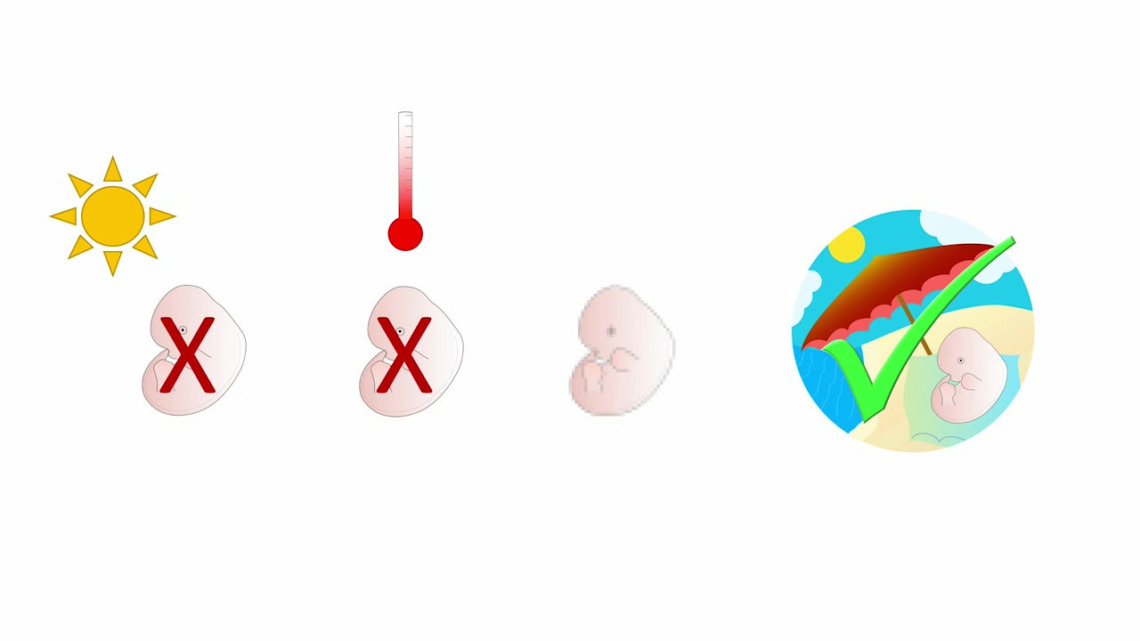
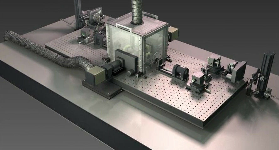
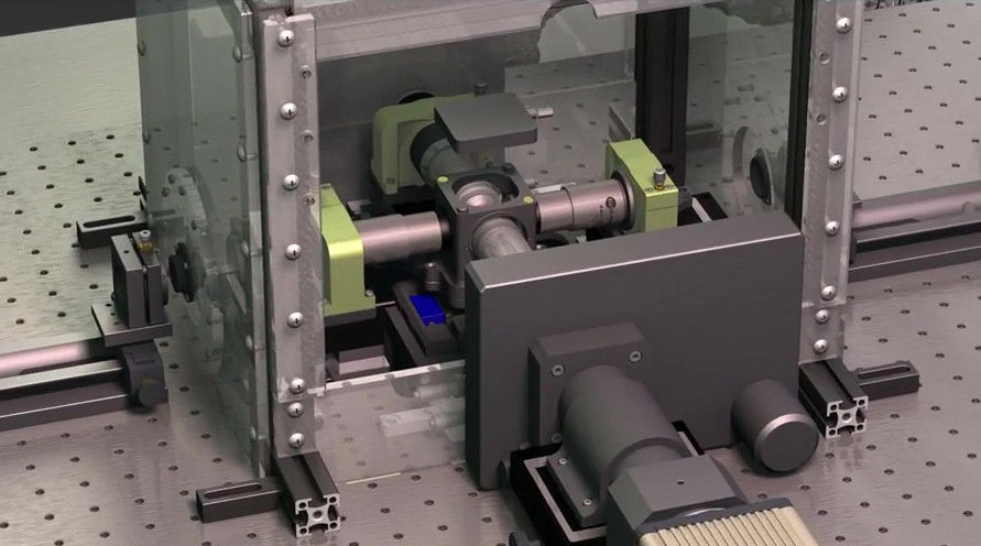
A special microscope called "Adaptive light-sheet microscopy" was developed to clear this difficult problem. Because mouse embryos are sensitive to light, it is possible to shut out light, sterilization processing is possible with precise control of atmospheric pressure and temperature.
In addition, there is a major advantage that a growth prediction algorithm incorporating image recognition technology by machine learning is adopted in order to keep pursuing a moving embryo at the cell level.
With the Adaptive light-sheet microscopy, you can check how the cell is organized in the following movie.
New Microscope Offers 4-D Look at Embryonic Development in Living Mice on Vimeo
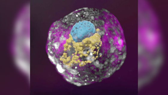
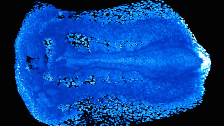
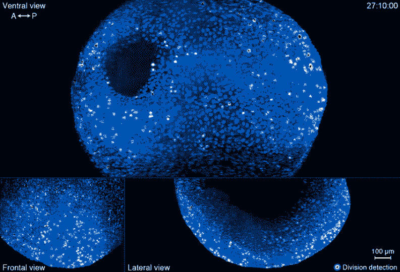
This is a mouse embryo imaged with Adaptive light-sheet microscopy. It is still in a single cell state and will continue to differentiate from it to an organ.
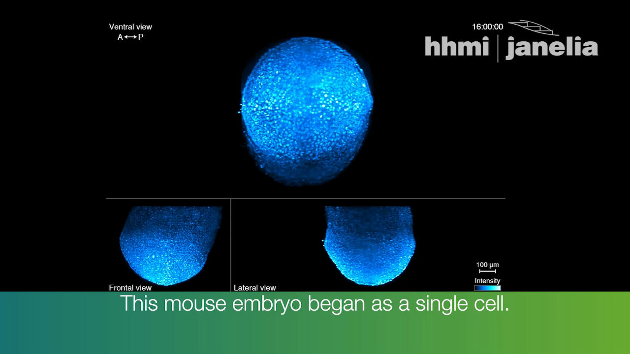
The image created with the Adaptive light-sheet microscopy is completely captured of the state of the embryo in which the cell continues to divide and grow. In addition, mouse embryos are said to be divided into 60,000 cells in less than one week
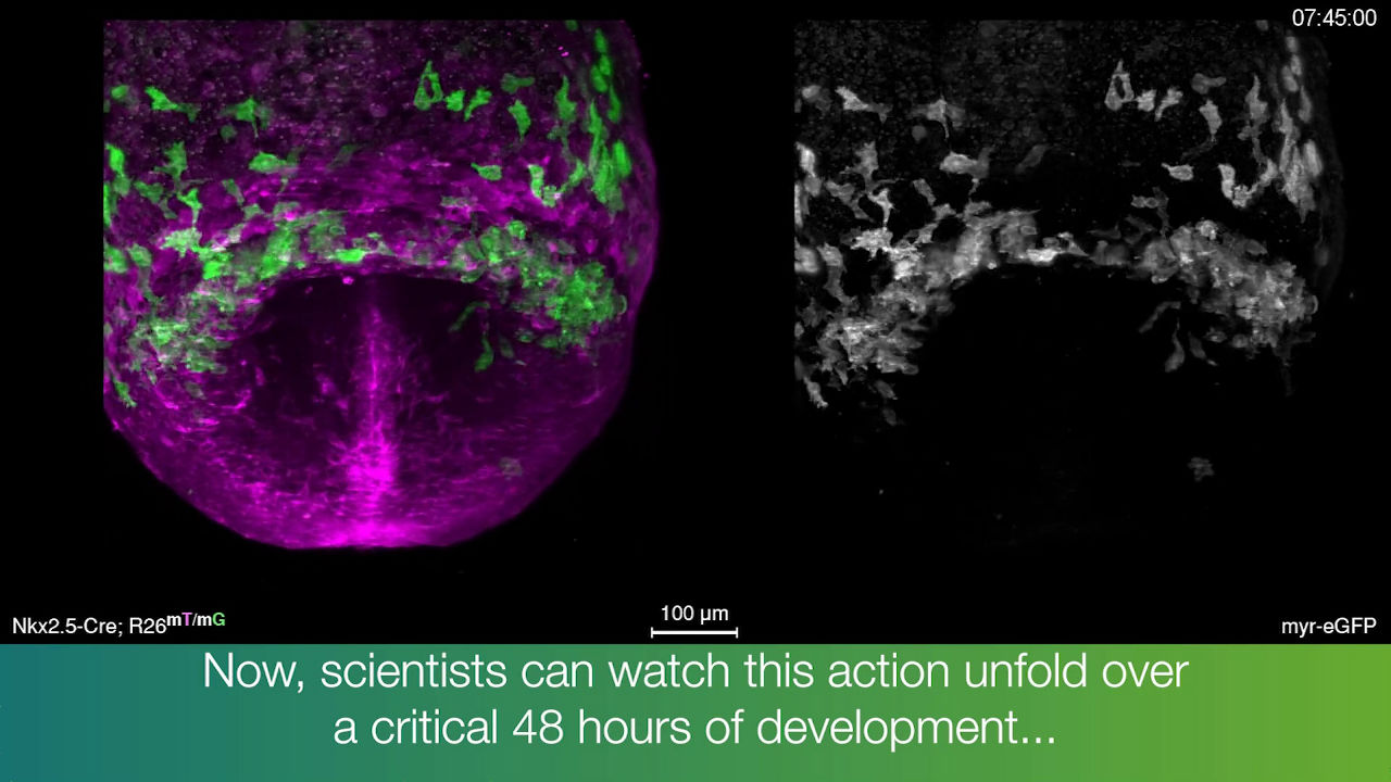
Successful to capture how the early heart starts beating heartbeat.
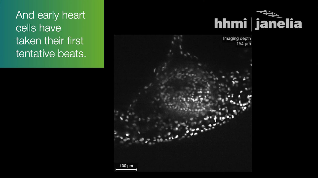
With the Adaptive light-sheet microscopy, it became possible to know by image the 48 hours most important for cell differentiation starting from 6 and a half to 8 and a half after fertilization.
In Adaptive light-sheet microscopy, two light-sheets illuminate the embryo and two cameras record one image every 10 milliseconds. Dr. Keller's research team has developed a calculation method called "TARDIS" to accurately track the size and position of the embryo. TARDIS, a calculation tool kit created from approximately 1 million images, quantifies the differences in individual embryonic development data by aligning the four embryos in space-time and space, so that the "average embryo" can be virtualized It was made in a way. "It would take a couple of years to track all the cells without TARDIS," Dr. Keller said to be an important technical element of the Adaptive light-sheet microscopy, as mentioned.
Howard Hughes Medical Institute is planning to allow Adaptive light-sheet microscopy to be used by other scientists. Adaptive light-sheet microscopy can be expected to further advance research on the mechanism of how cells evolve into organs.
Related Posts: