What is 'expansion microscopy,' which involves inflating cells with an absorbent material also used in disposable diapers, and then observing them?
If you want to see cells in detail under a microscope, you probably think you need more powerful and expensive equipment. 'Expansion Microscopy' overturns that notion. By using absorbent materials like those used in disposable diapers to enlarge cell and microbial samples, it becomes easier to see their fine structures under a standard microscope. Quanta Magazine, a science news site, has summarized the spread and use of expansion microscopy in microbial research.
Expansion Microscopy Has Transformed How We See the Cellular World | Quanta Magazine
Expansion microscopy - Wikipedia
https://ja.wikipedia.org/wiki/%E8%86%A8%E5%BC%B5%E9%A1%95%E5%BE%AE%E9%8F%A1%E6%B3%95
Expansion microscopy, developed by Ed Boyden and his colleagues at MIT in 2015, involves spreading a gel-like material that absorbs water and expands throughout the sample to be observed, physically enlarging the sample before observation. By attaching markers to the gel to maintain the relative positions of the structures of interest and then allowing the gel to absorb water, the distance between structures that were originally too close to distinguish increases, making it easier to distinguish subtle differences even with a conventional microscope. The material used to expand the sample is sodium acrylate, a water-absorbing material also used in disposable diapers.

Quanta Magazine explains that the unique feature of this technique is that even without an expensive, ultra-high-performance microscope, it is possible to improve the visibility by devising sample preparation. Omaya Dudan of the University of Geneva evaluates expansion microscopy as 'a low-cost, easy-to-learn technique that can produce better images even with inexpensive microscopes.'
'Dudin had struggled for many years to visualize the internal structure of objects with hard cell walls by passing antibodies into the cells to stain specific proteins. Previously, this was achieved using a complex process combining freezing and thawing, but in the end, not many samples were usable. However, when he tried expansion microscopy in a collaborative research project with a neighboring laboratory, he was able to successfully stain the samples in an expanded state, which dramatically advanced his observations,' Dudan said.
Expansion microscopy has also worked well in Gautam Dey's lab at the European Molecular Biology Laboratory. It has made it easier to see structures inside cells, and it's also easier to incorporate dyes and antibodies. As a result, Dudan's lab and Dey's lab have begun collaborating to examine a variety of microorganisms, making it easier to compare differences in cytoskeleton structures that were previously difficult to see.
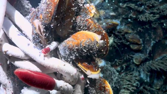
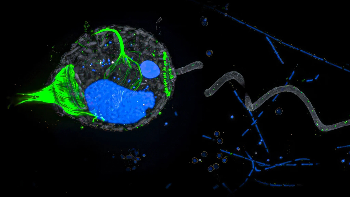
The image below shows the first image of a multi-nuclear protist,
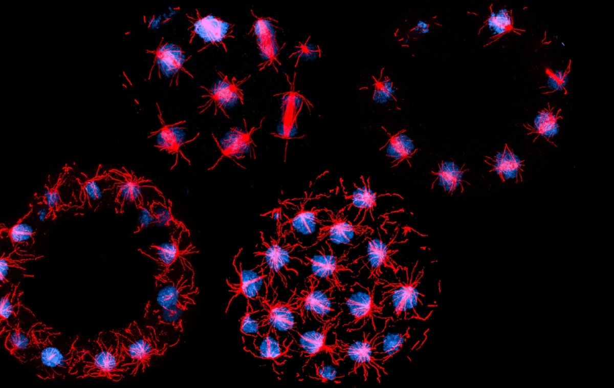
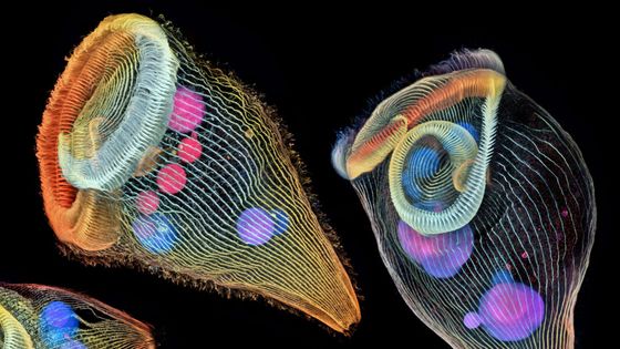
The predatory
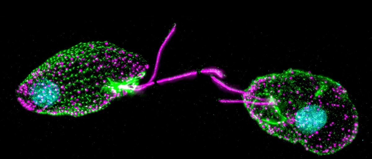
The image below shows Linomonas, a single-celled photosynthetic alga with two flagella found primarily in marine environments. Dudan and Day used expansion microscopy to observe a variety of unexpected structures, including the appearance of a special protein called centrin, which appears to frame the regularly arranged array. This had never been reported before in the Linomonas lineage.
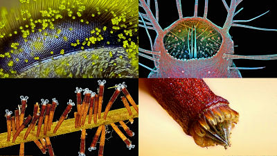
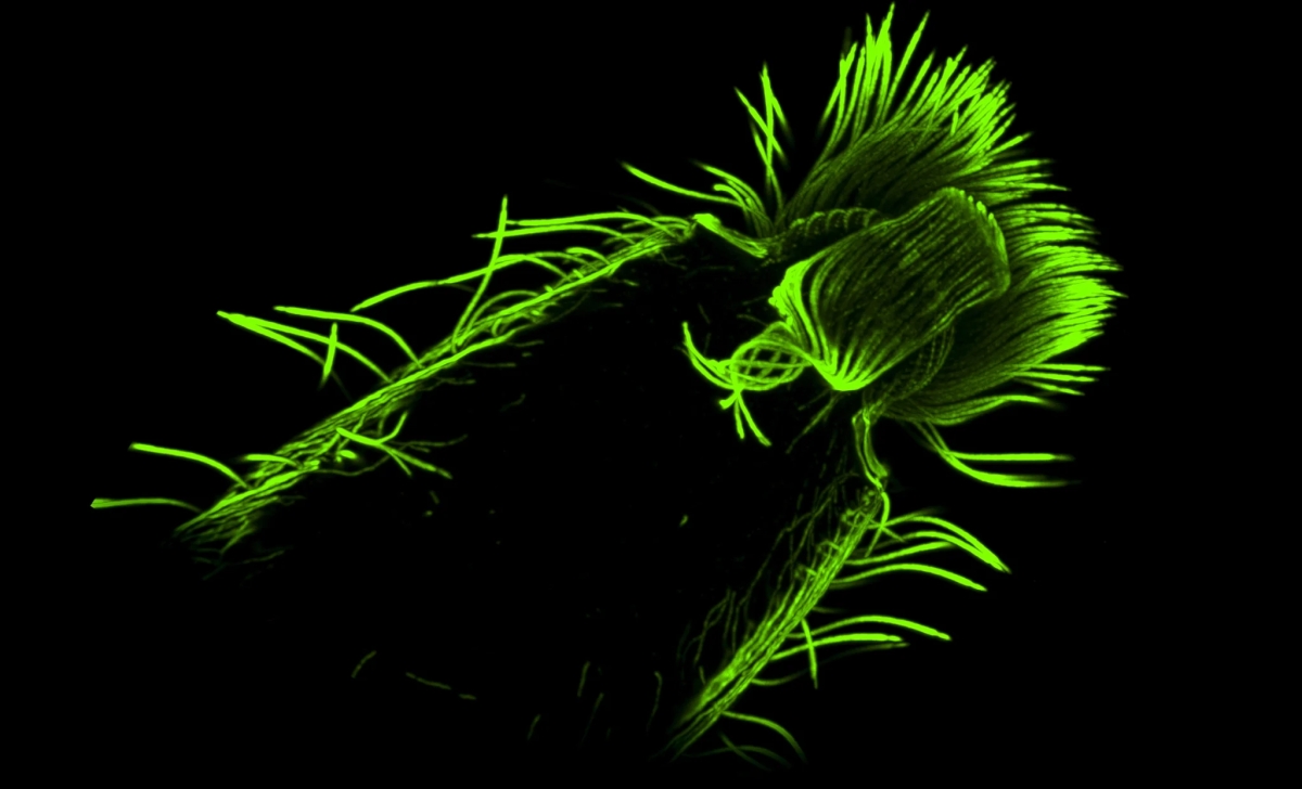
Diatoms are particularly difficult to image because they are covered in a glassy shell called a 'frustum.' Day's lab captured the images below using a different expansion microscopy protocol: 'We expanded the sample, tagged its proteins with fluorescent antibodies, and then flash-frozen the entire sample to fix it in place.'
While Quanta Magazine acknowledges that there are tricks to pre-processing the sample to expand it cleanly, and that adjustments to the conditions may be necessary depending on the subject, it points out that the great significance of this method lies in the fact that it can expand the world we can see by expanding the sample without relying solely on the performance of the device.
Related Posts: