It turns out that certain brain networks are on average 73% larger in people with depression than in healthy people

The human brain is made up of interconnected networks of different regions that carry out specific processes, and a new study comparing the brains of depressed and non-depressed people has found that certain brain networks are enlarged by an average of 73% in the brains of depressed people.
Frontostriatal salience network expansion in individuals in depression | Nature
Part of brain network much bigger in people with depression, scientists find | Depression | The Guardian
https://www.theguardian.com/society/article/2024/sep/04/part-of-brain-network-much-bigger-in-people-with-depression-scientists-find
In recent years, an approach called 'precision functional mapping,' which collects fMRI data from a large number of people to investigate inter-individual variations in brain networks, has attracted attention. However, precision functional mapping has not been used to clarify the differences between the brains of depressed and healthy people so far.
A research team from Cornell Medical School and other institutions conducted precision functional mapping on 141 depressed subjects and 37 non-depressed controls to precisely measure the size of brain networks and examine the average size of each.
They found that a brain network region called the frontostriatal salience network was on average 73% larger in people with depression than in healthy people.
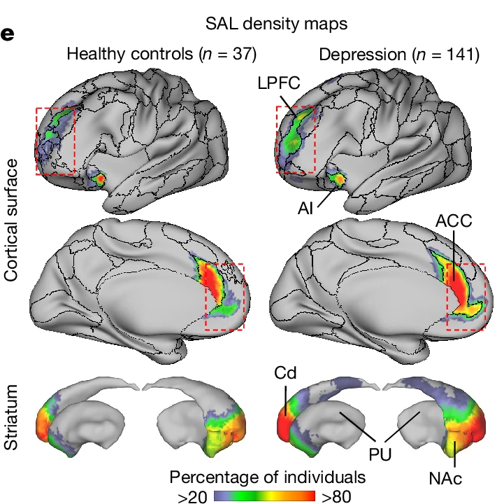
The following figure shows a mapping of the brain's salience network, with a healthy person on the left and a person with depression on the right. Depending on the proportion of people who had a salience network in a particular area, the lower the proportion, the more bluish the color, and the higher the proportion, the more reddish the color. Comparing the images, we can see that people with depression have a wider salience network in the lateral prefrontal cortex (LPFC), anterior cingulate cortex (ACC), caudate nucleus (Cd), putamen (PU), and nucleus accumbens (NAc).
These results were supported by an analysis of brain scans collected from 932 healthy subjects and 299 people with depression. The researchers found that the size of specific brain networks in the depressed subjects did not change with time, mood, or
However, when subjects experienced certain depressive symptoms, the synchronization of brain signals between different regions of the network weakened, and these changes were also associated with the severity of future depression.
In addition, an analysis of brain scan data from 57 children who developed depression during adolescence revealed that the brain networks that expanded in those with depression began expanding several years before symptoms appeared, and that similar expansion was observed in people who developed depression as adults.

These results suggest that the expansion of certain brain networks may be a risk factor for developing depression, rather than a consequence of depression. However, it is unclear whether the expansion of brain networks is caused by genetics or by certain experiences. It is also unclear whether the expansion of brain networks itself is related to depression, or whether the expansion of certain brain networks is associated with the shrinkage of other brain networks.
The research team said that this discovery could lead to the development of methods to examine the risk of developing depression in the future, as well as to the development of personalized treatments. Professor Connor Liston, co-author of the paper, commented, 'I think that the fact that we have identified a brain network that is related to depression and may be a risk for depression is reassuring for some people.'
Related Posts:


in Science, Posted by log1h_ik