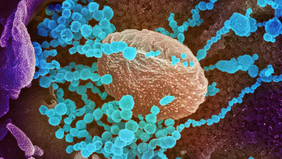
If you make it easy to understand the very moment of the new coronavirus infection and the replication mechanism with animation, it will be like this
The delta strain of the new coronavirus (SARS-CoV-2) is still rampant around the world, but much more information about the virus is available compared to the early epidemics of the new coronavirus infection (COVID-19). increase. Based on such information, biochemists and scientific animators from the University of Utah cooperated to animate 'the very moment when SARS-CoV-2 is infected'.
SARS-CoV-2 Visualization and Annotation Project — The Animation Lab
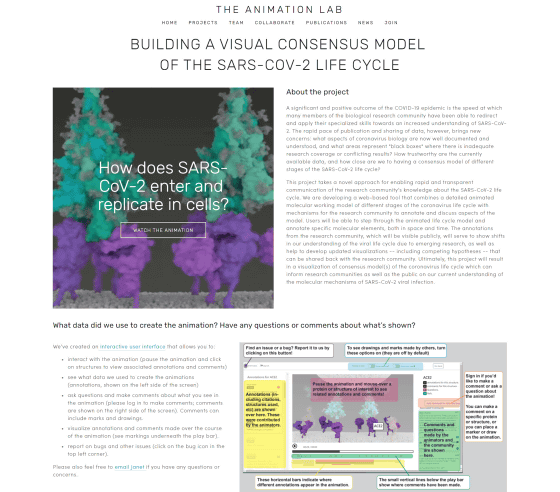
Coronavirus Annotation
https://coronavirus-annotation-3.sci.utah.edu/
How the coronavirus infects cells — and why Delta is so dangerous
https://www.nature.com/articles/d41586-021-02039-y
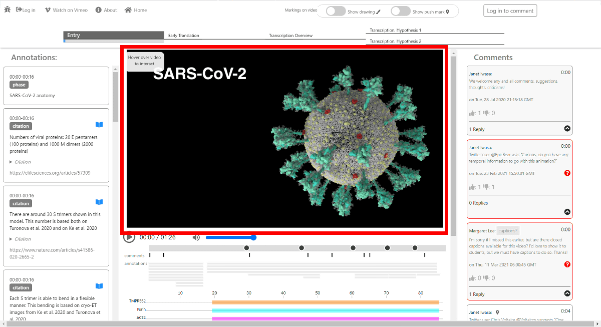
When you access 'Coronavirus Annotation', it looks like this. Click the image in the center of the screen to start the animation.
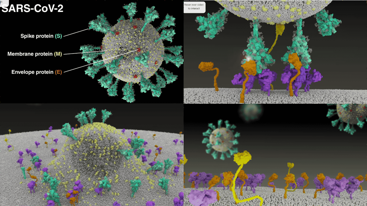
Of SARS-CoV-2, the
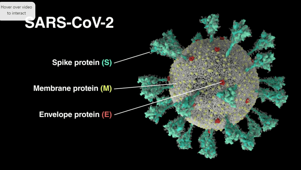
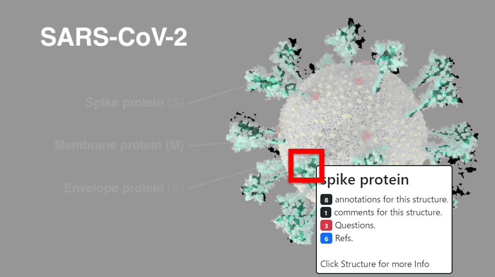
The animation is interactive, and the explanation of 'what it is' is shown in the part where the cursor is placed.
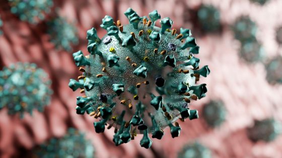
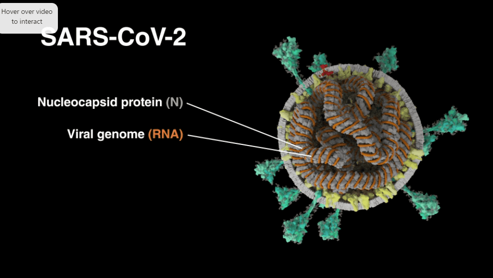
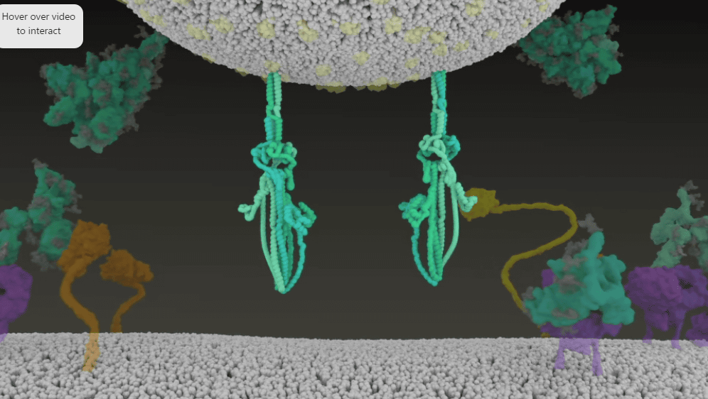
The cross section looks like this. The nucleocapsid protein is gray and the RNA is orange.

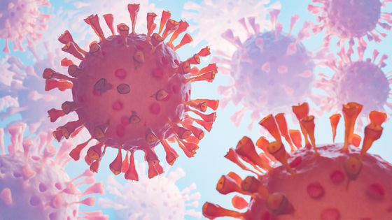
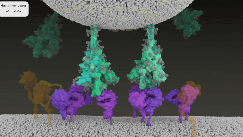
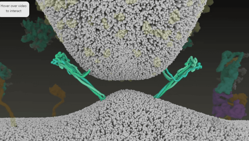
Influenza virus ectofusion proteins are relatively hard, but SARS-CoV-2 peaplomers are extremely flexible, allowing them to bend, sway, and rotate like hinges to facilitate binding. That thing. The unevenness of the spike protein matches the unevenness of ACE2 ...
Furin is also linked to membrane proteins.
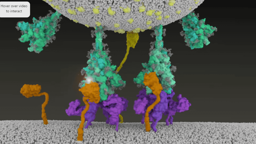
TMPRSS2 cuts the part of the pedplomer in which the body binding domain (RBD) is present.
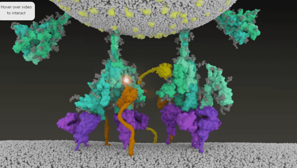
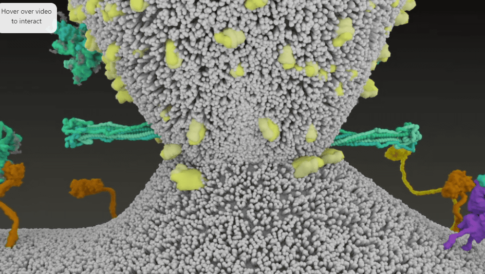
Then, the spike protein is decomposed, and ACE2 etc. take the parts ...
Hydrophobic amino acids flow out of the spike protein.
This sticks in the cell membrane ...

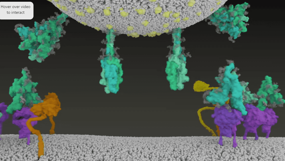
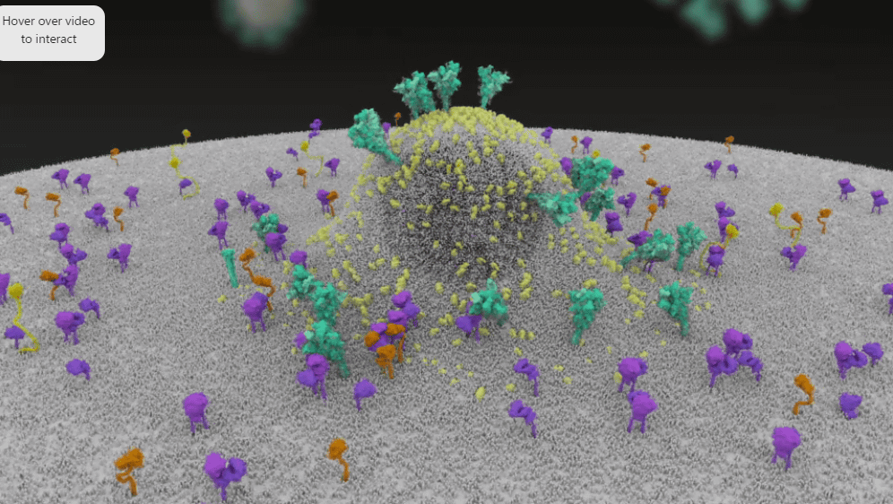
By bending tightly, the distance between SARS-CoV-2 and the cell membrane is reduced.
Virus and cell adhere.
Then, the viral genome is taken up into the cells.
There is a slider under the animation, you can play from the part you like, and under the slider it is easy to see when the color-coded protein etc. will appear in the animation.
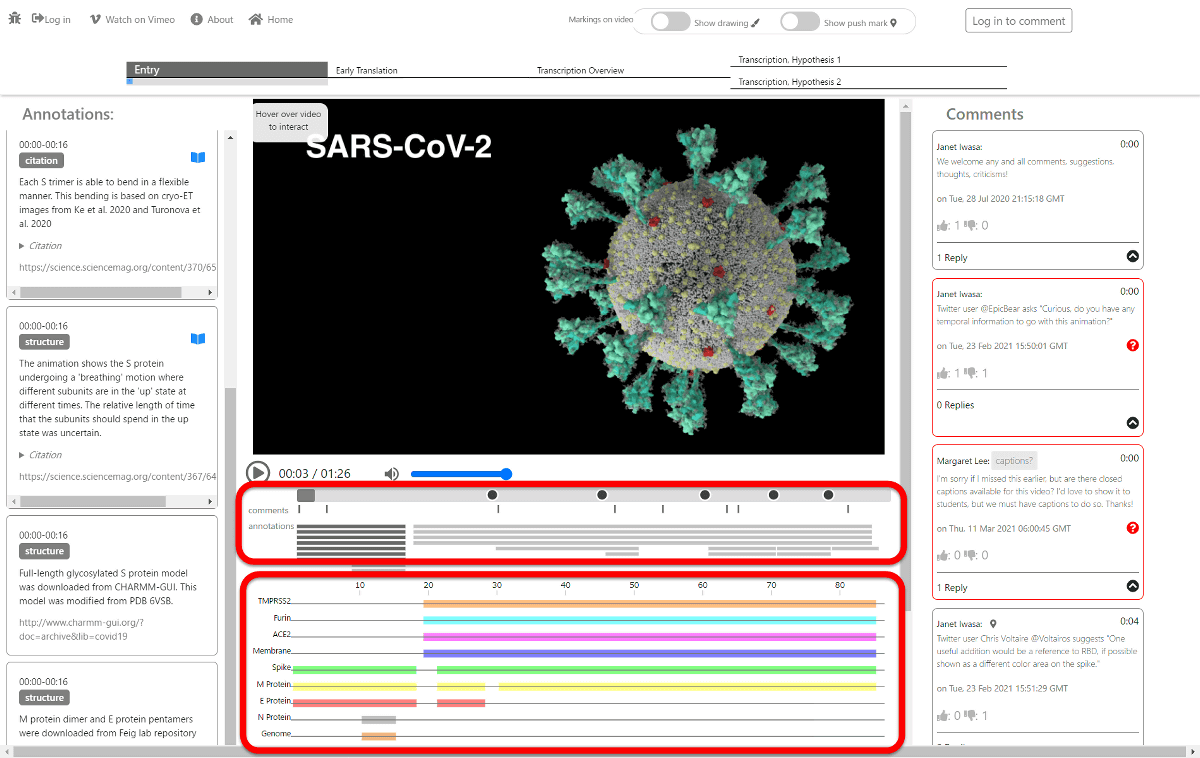
In addition, there is an annotation on the left side of the screen, for example, the part surrounded by the red frame describes what is happening at '00: 17-01: 24' on the timeline.

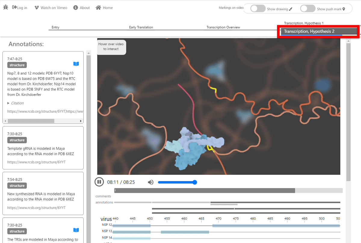
In addition, there is a tab at the top of the animation, and you can see the process of translating the protein by clicking 'Early Translation' next to the 'Entry' that animated the moment of infection.
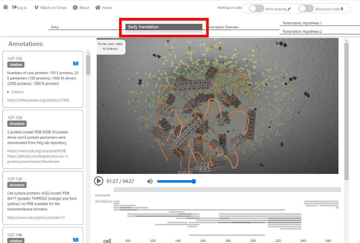
The tab next to it is 'Translation Overview'

In the tab on the far right, say 'Translation Hypothesis 1' ...
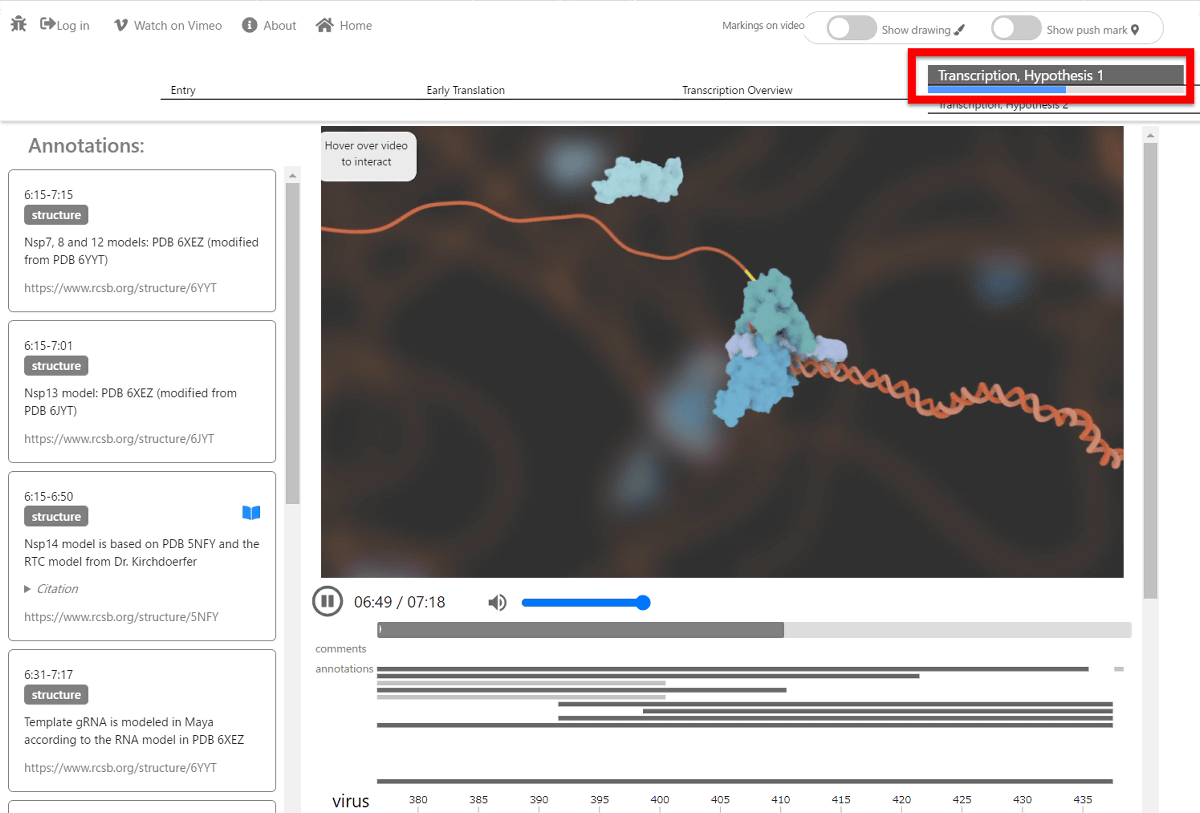
You can see 'Translation Hypothesis 2'.
Related Posts: