Award-winning works of art competition "BioArt" competing as a work of art in the world of nature and living things will be announced
Competition to evaluate microscopic world images and videos captured by microscope etc. as artworks "BioArt(Bio Art) "prize-winning work was announced. The fine world that we can not usually see is expressed vividly in color.
BioArt Image and Video Competition 2015 Winners Announced
http://www.faseb.org/Resources-for-the-Public/News-Room/Article-Detail-View/tabid/1014/ArticleId/1229/BioArt-Image-and-Video-Competition-2015-Winners-Announced.aspx
The colorful, dark, dynamic art of life: 2015 BioArt winners | Ars Technica
http://arstechnica.com/science/2015/11/the-colorful-dark-dynamic-art-of-life-2015-bioart-winners/
This competition is the AmericanFASEB(Federation of American Societies for Experimental Biology) held annually. The work won in 2015 has 10 images and 2 images.
FASEB> Resources for the Public> Scientific Contests> BioArt> 2015 BioArt Winners
http://www.faseb.org/Resources-for-the-Public/Scientific-Contests/BioArt/2015-BioArt-Winners.aspx
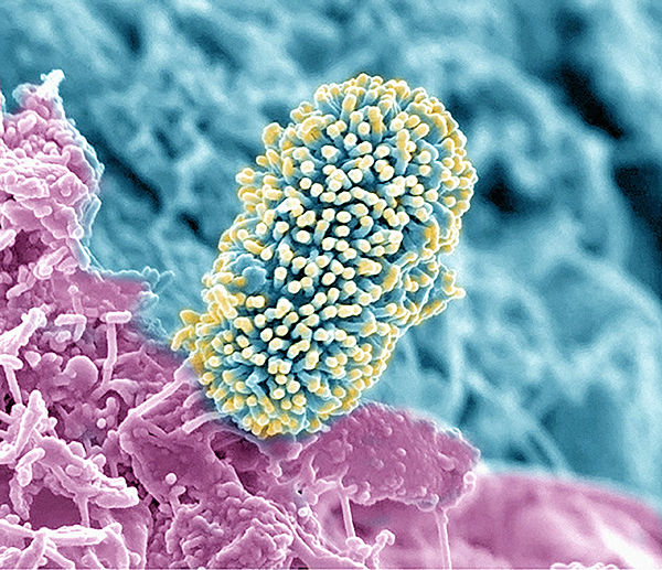
◆ Alice Dohnalkova(Environmental Molecular Sciences Laboratory, Pacific Northwest National Laboratory, Richland, WA)
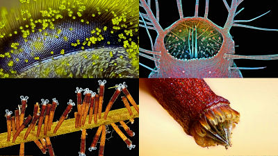
The state of the soil bacteria (yellow) which moved to the new residence by moving the surface (Arabidopsis thaliana root surface (purple · blue)) was taken with a scanning electron microscope. In the research, it is investigated how the availability of carbon around the root affects the habitat of bacteria, and it is expected that it will be useful for the method of generating bioenergy without affecting the environment in the future It is said that.
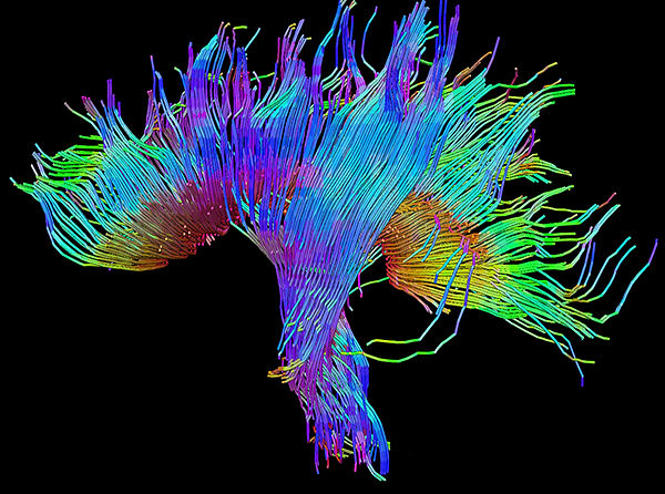
◆ Xiawei Ou(Arkansas Children's Nutrition Center, Arkansas Children's Hospital, and University of Arkansas for Medical Sciences, Little Rock, AR)
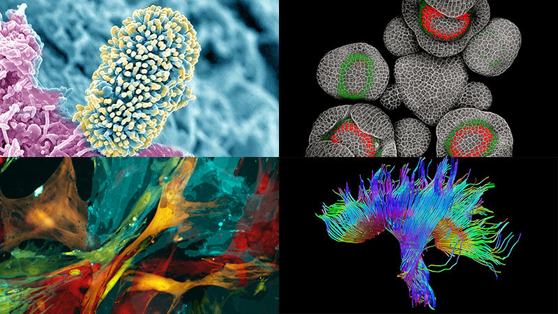
Nerve fibers connecting the region of the human brainDiffusion tensor imagingMapped using 3D mapping. What is reflected hereCorticospinal tractWhenCorpus callosumThe bundle of nerve fibers, color from left to right red, from front to green, from top to bottom blue, color-coded according to the direction of the fiber.
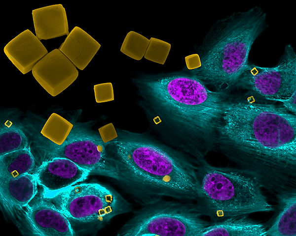
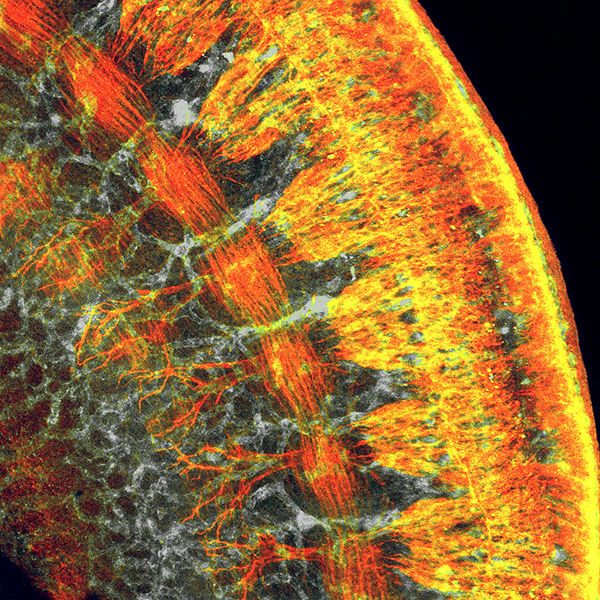
◆ Jenolyn F. Alexander(Houston Methodist Research Institute, Houston, TX),Veronika Kozlovskaya,Eugenia Kharlampieva(University of Alabama at Birmingham, Birmingham, AL),Biana Godin(Houston Methodist Research Institute, Houston, TX)
This image was created by putting anticancer drug in cube-shaped micro packaging container and delivering it directly to human breast cancer cells to synthesize how to use for treatment. Research using scanning electron microscopy and confocal fluorescence microscopy is progressing on how microparticles of various sizes and shapes can deliver drugs pinpoint to specific cells.
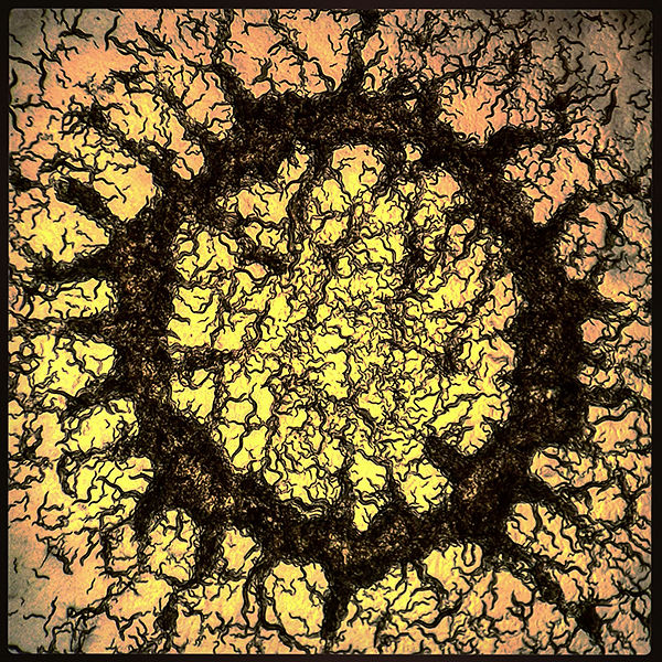
◆ Adam Brown,David Biron(University of Chicago, Chicago, IL)
Caenorabditis eleganceCalled nematodecolony(Aggregation). Ring-shaped colonies are formed along the state that many food bacteria are hardened. Caenorabditis elegans with a simple nervous system are used to study the influence of serotonin, which is a neurotransmitter in the brain, and the feeding behavior.
◆ Heinz Baumann, Sean T. Glenn, Mary Kay Ellsworth, Kenneth W. Gross(Roswell Park Cancer Institute, Buffalo, NY)
A method of tracking cells by coloring cells in various colors, and taking pictures of mouse pancreatic cells. By coloring cancer cells, it becomes possible to distinguish from the original organ and observe how the cancer cells diffuse into the body.
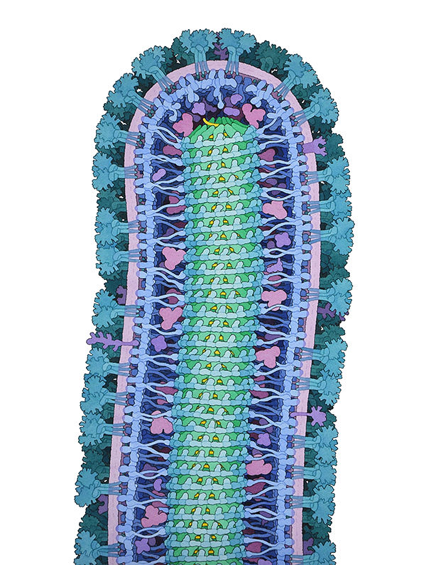
◆ David S. Goodsell(Research Collaboratory for Structural Bioinformatics Protein Data Bank, Piscataway, NJ / La Jolla, CA)
An illustration of the complex structure of Ebola cells. Only the compact RNA genome shown in yellow has the role of giving instructions to seven kinds of structural proteins indicated by blue, green, magenta and so on, and it is a mechanism for making activities as a virus.
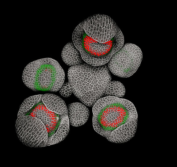
◆ Nathanaёl Prunet, Elliot Meyerowitz, Thomas JackCalifornia Institute of Technology, Pasadena, CA, Dartmouth College, Hanover, NH, Howard Hughes Medical Institute)
A picture of a bud of Shirona yona. Activating the "SUPERMAN gene" shown in red works to prevent stamens and pistils from interfering with each other.
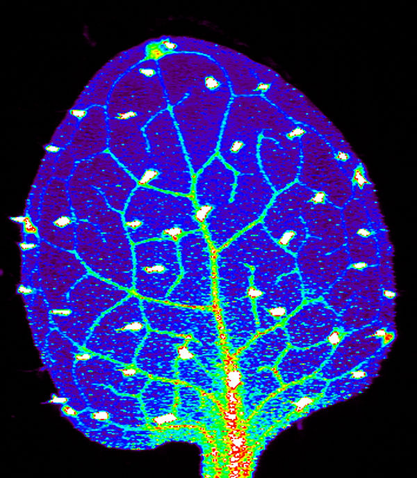
◆ Suzana Car, Maria Hindt, Tracy Punshon, Mary Lou Guerinot(Dartmouth College, Hanover, NH)
Expanded leaves of Shiroyu ni zoznaa using synchrotron X-ray fluorescence technique that tracks the micronutrient zinc, which greatly affects human health by helping human brain growth and immune system function properly. A study to improve the content of nutrients in crops by studying the state where plants accumulate specific substances.
◆ Shachi Bhatt Paul Trainor(Stowers Institute for Medical Research, Kansas City, MO)
By observing the mouse fetal cells in detail, we investigated the formation of a structure in which blood vessels (ash) and nerve cells (red) are parallel. It is useful for research on fetal birth defects and developmental disorders.
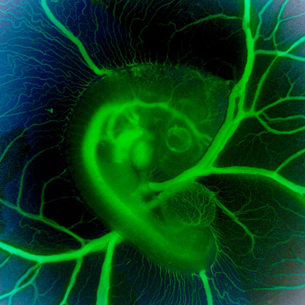
◆ Jessica Ryvlin, Stephanie Lindsey, Jonathan Butcher(Cornell University, Ithaca, NY)
A photograph of a state in which the heart of the fetus of the chick was subjected to microscopic surgery and influenced blood flow. We observe changes in blood flow during the subsequent growth process and are studying disorders in the human heart.
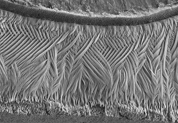
◆ Olivier Duverger, Maria I. Morasso(National Institute of Arthritis and Musculoskeletal and Skin Diseases, National Institutes of Health, Bethesda, MD)
This picture which looks like a fabric is a photograph of tooth enamel which is said to be the hardest in the human body using a scanning electron microscope.Enamel trabeculaeAre woven in a lattice pattern, a tough and flexible substance is formed.
[Winning work · movie section]
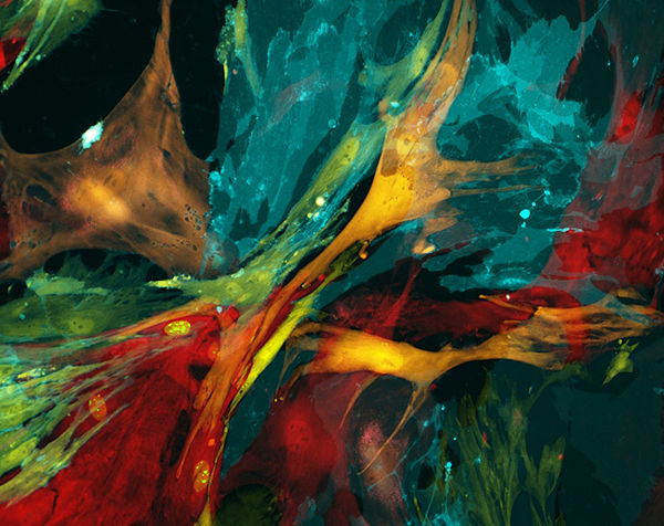
◆ Mehmet Berkmen and Maria Penil(New England BioLabs, Ipswich, MA)
In 2015, two movies are elected. The following movie is a long-time observation of the state of bacteria that makes flowers bloom on an agar plate. Cultivated bacteria such as Serratia (red), Bacillus (white), Nesserenrenia genus (yellow) and painted like a painting.
2015 BioArt Competition Winners: Mehmet Berkmen and Maria Penil - YouTube
◆ Kimberly Leiken, Elana Harris(Cincinnati Children's Hospital Medical Center, Cincinnati, OH)
A motion picture of the brain activity of a person with obsessive compulsive disorder measured using a helmet type magnetoencephalograph. The higher the level of activity in each area of the brain, the more red it is, the lower the activity level, the more blue it is shown. By visualizing the activity of the brain, it is expected to be useful for evaluating the patient's reaction response.
2015 BioArt Competition Winners: Kimberly Leiken and Elana Harris - YouTube
Related Posts: