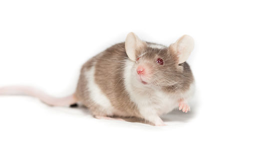
We successfully transplanted human brain cells into the brains of mice.
A research team from Stanford University and other institutions transplanted brain tissue made from human stem cells into mice lacking a large portion of their cerebral cortex and confirmed that it connected extensively with the mice's nervous system. The transplanted tissue grew, extending nerve fibers to the spinal cord, and showed changes in some memory tasks and gait after hypoxic injury. The research findings were published in Nature on September 16, 2026, and are reported as a new experimental model for studying human brain development and disease.
Developmental xenocortication using human-derived organoids in mice | Nature
Human brain cells transplanted into mice in 'most extensive' integration ever | Nature
https://www.nature.com/articles/d41586-026-02912-8
Because opportunities to experimentally examine living human brain tissue are limited, researchers have used 'brain organoids,' which are stem cells cultured in three dimensions, to study brain development and disease mechanisms. However, organoids in culture dishes lack blood vessels and an environment for exchanging signals with the body. Therefore, there are limitations to studying nerve cell maturation and the influence of neural circuits on behavior.
In 2022, neuroscientist Sergiu Paschka and his colleagues at Stanford University demonstrated that transplanting human brain organoids into newborn rats allows nerve cells to mature and connect to sensory neural circuits. In 2024, they used this method to evaluate potential treatments for Timothy syndrome , a severe genetic disorder associated with autism and epilepsy.
In conventional transplants, human tissue is inserted between the rat's own brain tissue, which limits the space available for growth. Furthermore, because rat nerve cells develop faster than human nerve cells, by the time the human cells extend their nerve fibers, rat neural circuits have already formed around them.
In this study, to reduce competition for space and neural circuits, we created mice in which the cells forming the neocortex and hippocampus were significantly reduced in the early stages of development through genetic engineering. These mice had a total brain tissue volume that was about 50% smaller than the control group, but with modified rearing conditions, they survived to adulthood and retained basic mobility.
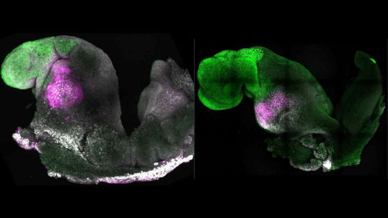
The research team transplanted four cerebral cortical organoids, created from human iPS cells, into the brains of newborn mice. The transplant success rate was 86.2% in the 29 mice examined, and the volume of the transplanted tissue increased approximately 4.7 times between two and three months after transplantation. At three months, the human-derived transplanted tissue accounted for 91.9% of the combined volume of the remaining mouse-derived cortical tissue and the transplanted tissue.
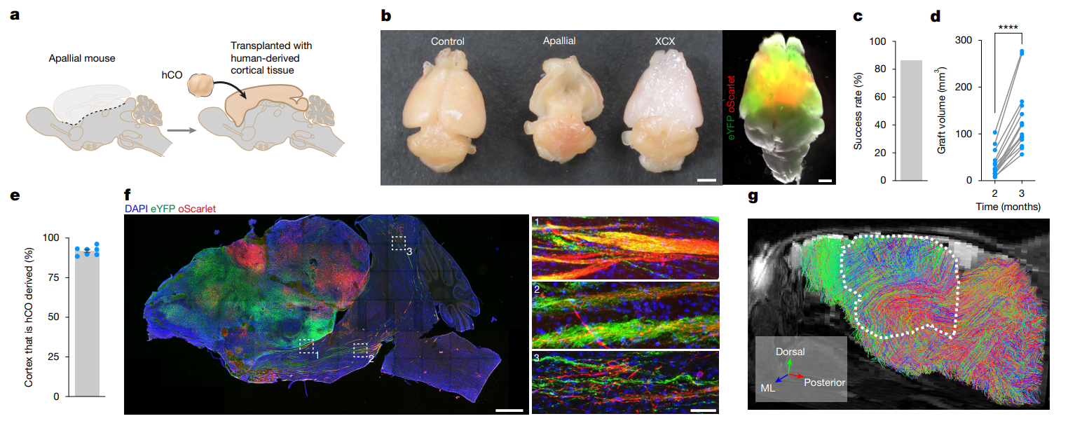
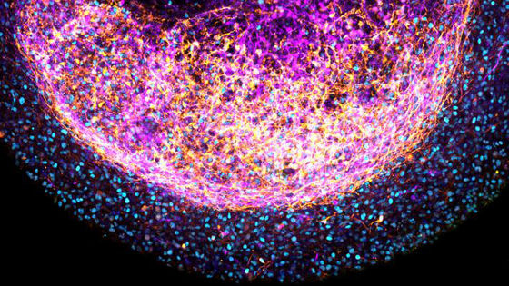
The transplanted tissue not only grew larger, but also extended nerve fibers to various parts of the mouse brain, some even reaching the spinal cord. Conversely, neural connections from the mouse thalamus and other areas to the transplanted tissue were also confirmed. Measurements of neural activity revealed patterns of synchronized cell activity, similar to developing neural circuits.
The transplanted tissue also produced a variety of cells that make up the human cerebral cortex. Among these were elongated nerve cells similar in shape to 'von Economo neurons,' large nerve cells that have been implicated in social cognition. These cells are difficult to reproduce in culture dishes and have been linked to frontotemporal dementia, so they may provide clues to investigating susceptibility to disease.
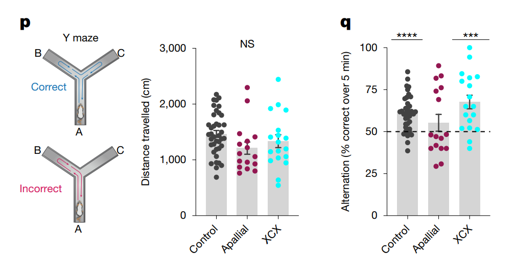
Behavioral tests showed that transplantation compensated for some functions. In a Y-shaped maze task that tested whether mice could remember the location they had explored most recently, untransplanted mice (reddish-purple) with significantly reduced cortex did not perform above the level of chance, while transplanted mice (light blue) and the control group (gray) performed above that level.
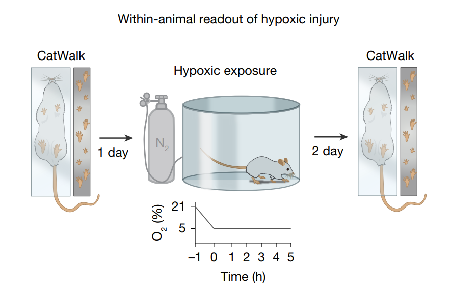
To further investigate its potential for disease research, the research team also examined damage caused by hypoxia, anticipating studies on oxygen deficiency before and after birth and related cerebral palsy. They found that transplanted tissue exposed to hypoxia showed an increase in oxygen-deficient-responsive proteins and cellular responses similar to those seen in brain injury.
Furthermore, when examining the gait of mice after exposure to hypoxia, the transplanted mice showed an increased proportion of simultaneous use of three to four paws to support their bodies. There was no change in overall running speed or gait, and the change is attributed to an alteration in leg coordination. The research team is focusing on the fact that damage to human-derived brain tissue can be evaluated not only in terms of cellular changes but also in terms of animal behavior.
It should be noted that this transplanted tissue does not replicate the mature human cerebral cortex. Clear layering and regional differentiation are incomplete, and the neural circuits are in the early stages of development. Furthermore, further investigation is needed to determine the extent to which the transplanted neurons are directly involved in specific behaviors, and whether synchronized activity within the transplanted tissue is transmitted externally.
Furthermore, research involving the transplantation of human brain tissue into animals raises ethical challenges. According to the journal Nature, which published the paper, the study underwent oversight, including independent bioethical review, and did not show any increase in the intelligence of the mice in behavioral tests. However, it is noted that further consideration will be necessary if more mature tissues are to be created in the future, or if animals with developmental rates closer to those of humans are used.
Related Posts:




in Science, Posted by log1i_yk