People with aphantasia, who are unable to form images in their heads, have different brain wiring

Imageless imagery in aphantasia revealed by early visual cortex decoding: Current Biology
https://www.cell.com/current-biology/abstract/S0960-9822(24)01652-X
People who can't 'see with their mind's eye' have different wiring in the brain | Live Science
https://www.livescience.com/health/neuroscience/people-who-cant-see-with-their-minds-eye-have-different-wiring-in-the-brain

The symptoms of aphantasia were first described in a statistical study in 1880 on mental imagery, which is the process by which people imagine objects or scenes. Professor Adam Zeman, a neurologist at the University of Exeter, began his research on mental imagery in 2005 and has spent the last decade listening to the stories of more than 12,000 people who describe themselves as aphantasia.
'As far as I see it, aphantasia is an interesting variation in the human experience, not a disorder,' Professor Zeman said of the condition.
The history of research into 'aphantasia,' the inability to visualize an image in one's head, and 'hyperphantasia,' the ability to instantly visualize a vivid image - GIGAZINE

A paper published in the journal Current Biology on January 10, 2025, looked at brain signals to understand why aphantasia occurs and what it means when you can't think of any mental imagery at all.
The study recruited 14 aphantasia patients and 18 non-aphantasia participants and conducted a test called 'binocular rivalry.' The brain constantly integrates visual information from the left and right eyes to build a single cohesive image. Binocular rivalry uses this property to flash two stripes of different colors in front of the subjects' eyes and ask them to answer which of the two stripes they perceived.
The results showed that people without aphantasia were more likely to be biased in which of the two patterns they recognized first, whereas people with aphantasia were much less likely to experience this bias. 'The stronger the mental imagery, the more likely it is that we will be biased in our perception of the binocular rivalry pattern,' explained Joel Pearson, a professor of psychology at the University of New South Wales and co-author of the study.

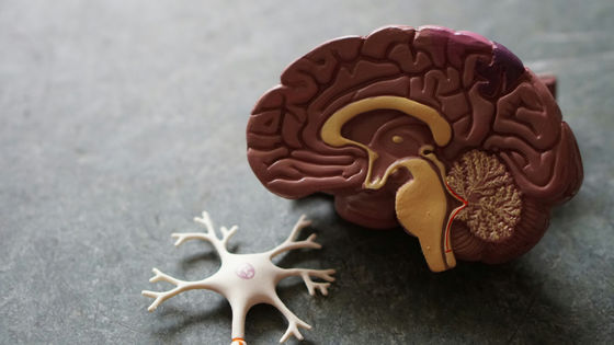
After testing for aphantasia with a binocular rivalry test, participants were given a similar stripe perception test using functional MRI, which tracks the flow of oxygenated blood, to study brain activity. The results showed evidence that both aphantasia and non-aphantasia participants had active primary visual cortex, the main part of the brain responsible for processing visual information, when looking at stripes and when imagining what the stripes were like, generating mental images. However, aphantasia participants showed slightly weaker activity in the primary visual cortex, suggesting that the brain may be performing a lower level or different type of processing when viewing images.
Furthermore, the brain activity was analyzed using a computer algorithm that was able to accurately predict the visual patterns that the subjects were trying to perceive or imagine. The results showed that perceptual and imaginative signals were clear in both subjects with and without aphantasia. However, while non-aphantastic subjects were able to roughly match what they perceived with what they imagined, in the aphantastic subjects the 'brain activity elicited by perception' and 'brain activity elicited by imagination' did not match, suggesting that fundamentally different processes may be occurring.
Pearson also said that a series of tests led to a surprising discovery. Normally, people process what they see in the right visual field with the left hemisphere, and what they see in the left visual field with the right hemisphere. However, people with aphantasia tend to process what they see with their right eye with the right hemisphere, and what they see with their left eye with the left hemisphere. 'It's possible that their brains are wired completely differently,' Pearson said.
Commenting on Pearson's research, Nadine Dijkstra, a senior research fellow at University College London, said: 'This study adds to the evidence that people with aphantasia use the visual cortex in a different way to people without aphantasia when imagining things. However, it is important to note that this study is small in scale and somewhat inconsistent with previous research in this field. Brain activity in people with aphantasia is a new area of research, and many questions remain to be answered.'
Related Posts:


in Science, Posted by log1e_dh