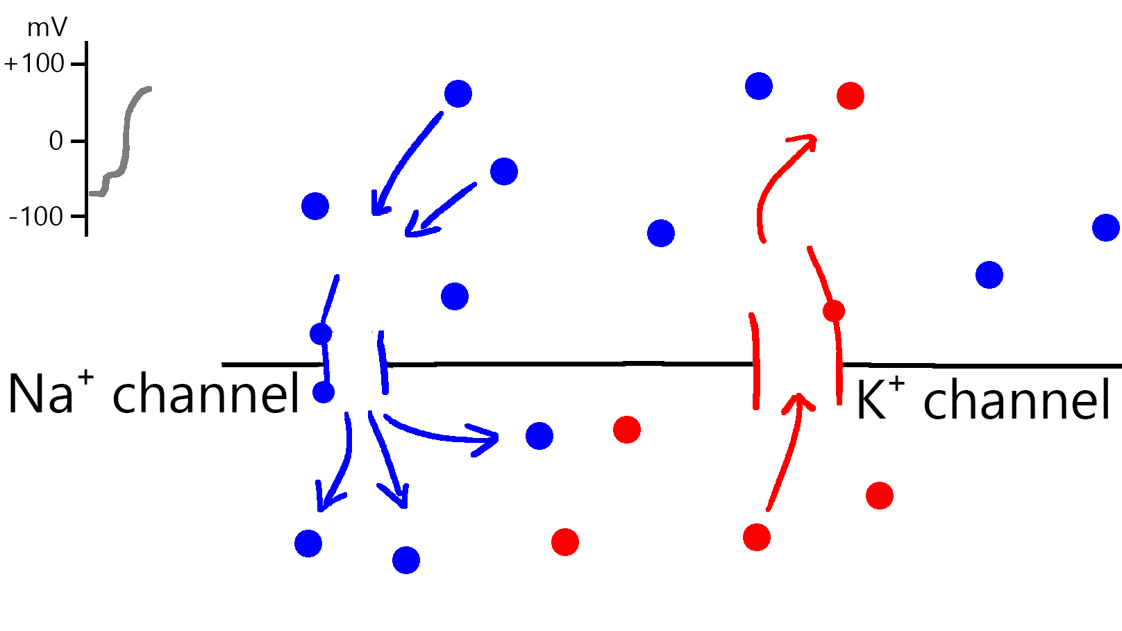
A rough illustration of the mechanism by which the ion channels of neurons regulate the potential difference looks like this
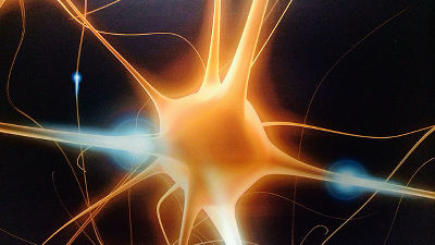
The cells that make up the brain, the mechanisms of
The magic of ion channels in the neurons |
https://i-kh.net/2020/08/26/the-magic-of-ion-channels/
Kramtsov explains the mechanism of ion channels in neurons as follows. Mr. Kramtsov's commentary is mainly quoted from the book ' Fundamental Neuroscience' .
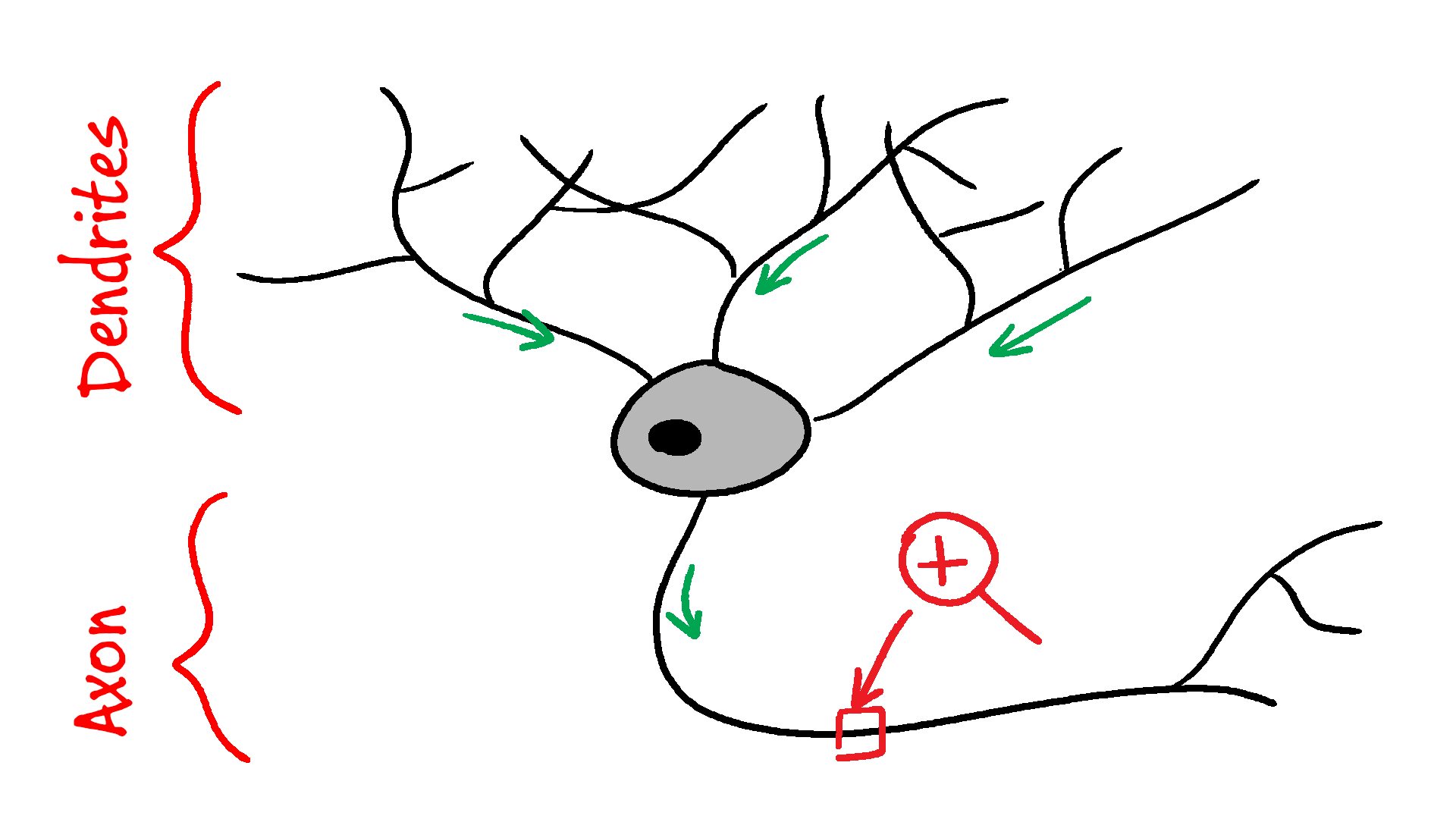
First, neurons are composed of cell bodies , dendrites, and axons . Multiple dendrites branching from the cell body are responsible for receiving signals from other cells, and axons are responsible for transmitting signals to other cells.
The cell bodies of neurons are covered with ions such as calcium, potassium, sodium, and chlorine. Ion channels on the surface of neurons are like tiny valves that allow ions to pass when certain conditions are met. Some ion channels open when they bind to certain chemical substances such as neurotransmitters, and others open when the intracellular potential difference increases. Ion channels that open when the potential difference increases are called 'voltage-gated ion channels', and there are channels corresponding to each ion such as sodium, potassium, and calcium.
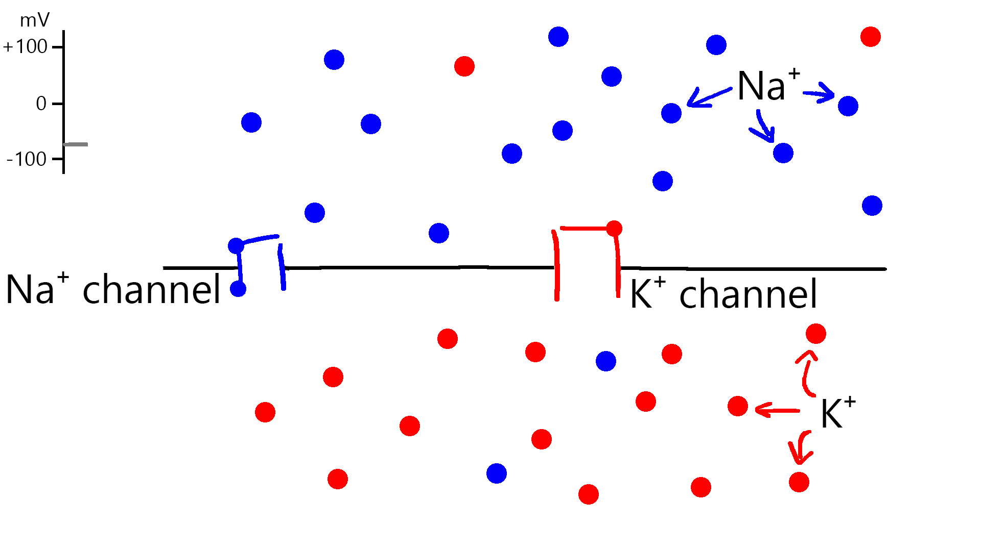
The figure below shows the activity of an ion channel at steady state. Blue dots are sodium, blue lines are voltage-gated sodium channels, red dots are potassium and red lines are voltage-gated potassium channels. The top of the central black line represents the outside of the neuron, and the bottom represents the inside of the neuron. The upper left is a graph showing the potential difference in the cell. The stable potential difference is around -70mV, and both channels are closed.
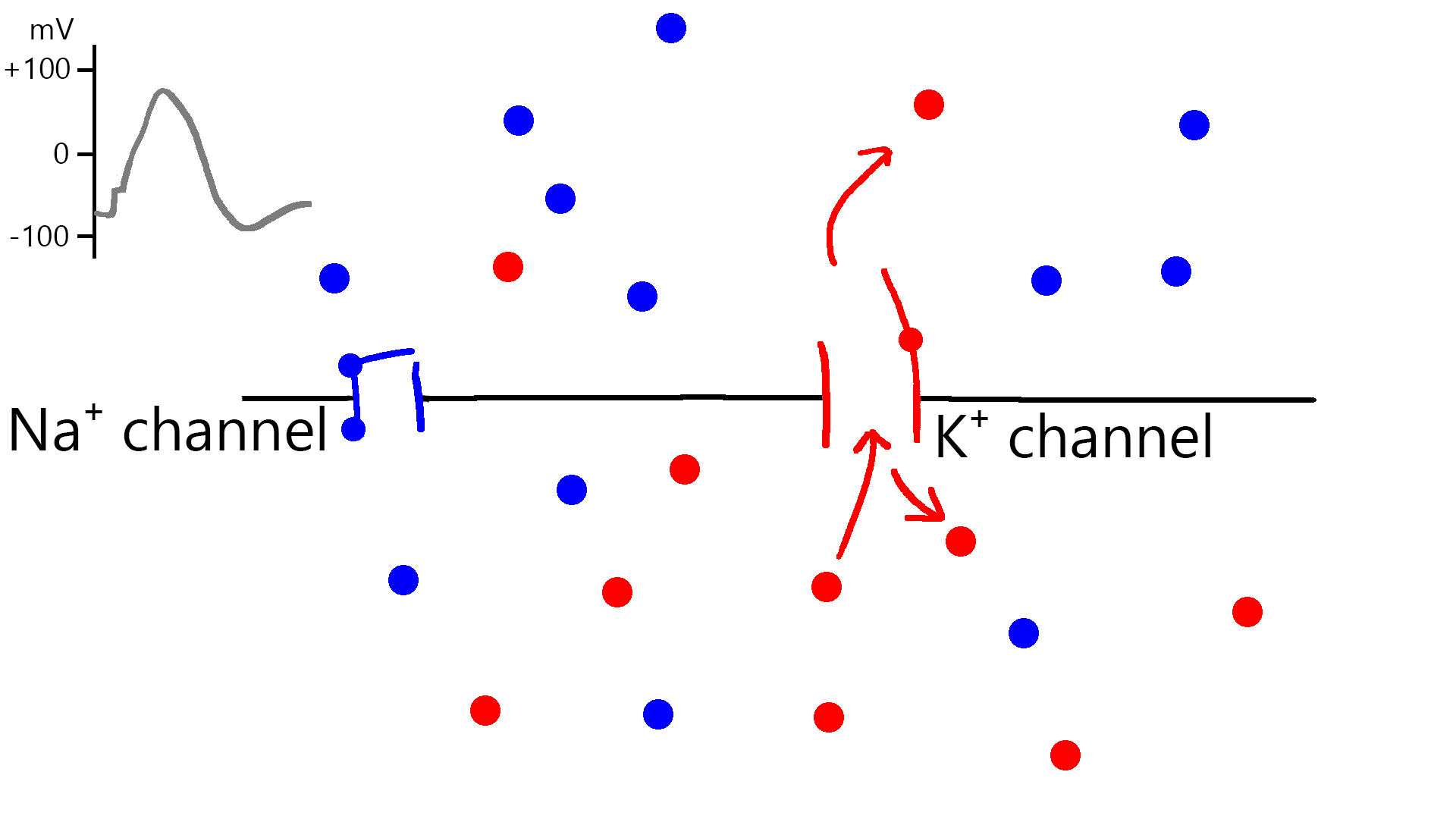
However, when the potential difference between inside and outside the neuron exceeds -55mV for some reason, the sodium channel opens and a large amount of sodium ions enter the neuron. Since the captured sodium ions have a positive charge, the voltage inside the neuron rises to +30mV. Potassium channels then also open, releasing several potassium ions against the positive charge of sodium, slowing the rise in potential difference.

After the sodium channel closes, the potential difference decreases by releasing potassium ions, and when the potential difference approaches a steady state of -70 mV, the potassium channel gradually begins to close. Since each channel opens and closes over a span of a few milliseconds, and the total number of ions taken in and released through each process is small, the overall concentration of ions inside and outside the neuron is largely unaffected. .
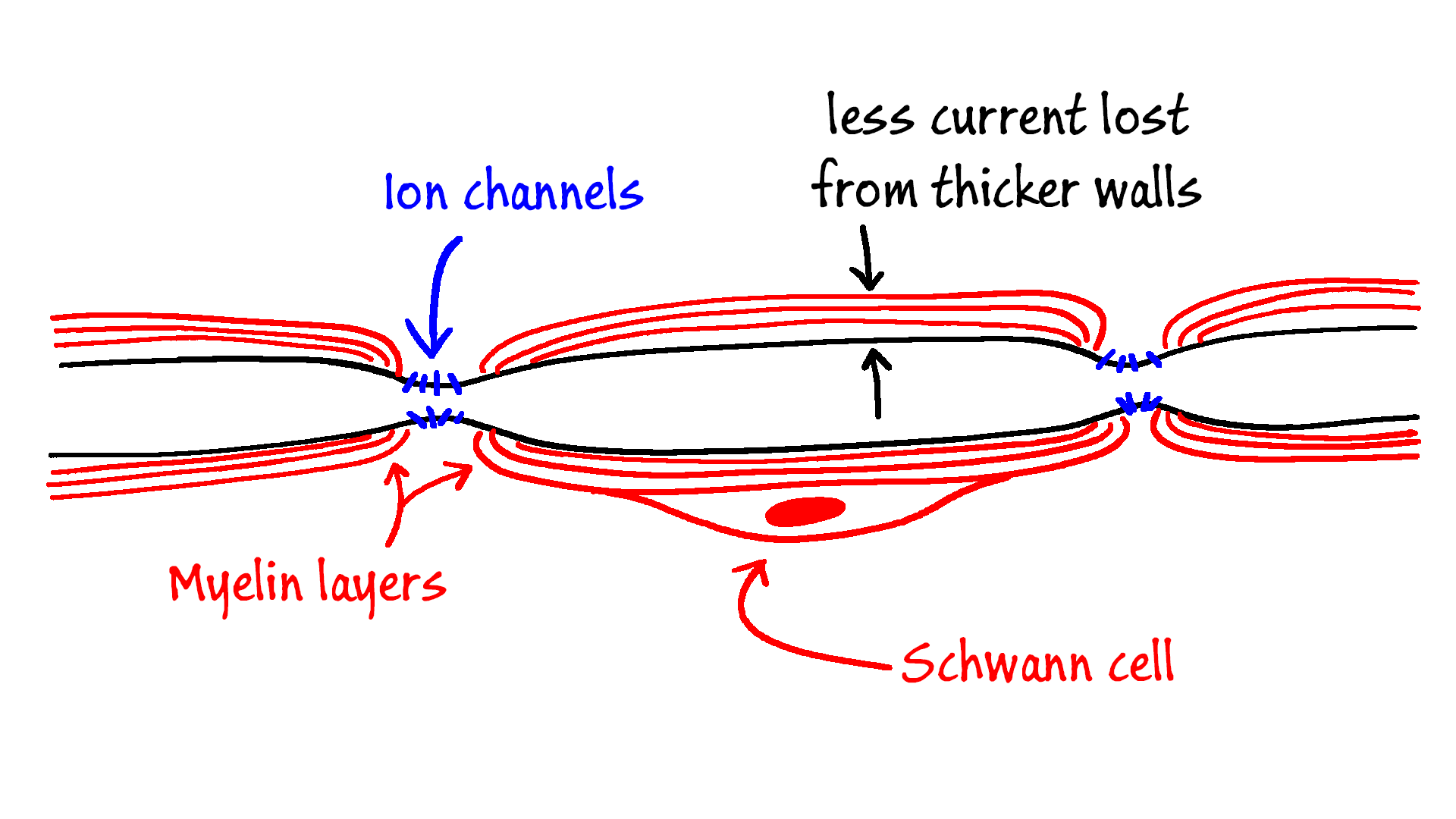
Axonal ion channels are evenly spaced. Ion channels are present in the myelin layers within neurons, which are not covered with insulator-like substances that block signal transmission between axons.

The myelin sheath has
Related Posts: