An attempt to convert 3D data from an elaborate human model made of ivory over 300 years ago
The human body model has a long history, and in Europe from the late 17th century to the beginning of the 18th century, models based on anatomical drawings of the human body were made of wood and ivory. A research team at
RSNA 2019-Radiological Society of North America Annual Meeting | HealthManagement.org
https://healthmanagement.org/c/imaging/event/rsna-2019-radiological-society-of-north-america-annual-meeting
CT scans confirm 17th-century medical mannikins are mostly made of ivory | Ars Technica
https://arstechnica.com/science/2019/11/micro-ct-scans-reveal-the-secrets-of-17th-century-anatomical-manikins/
The research team at Duke University has released a movie showing the human body model being scanned.
Doctor Dolls, Coming Soon in 3-D-YouTube
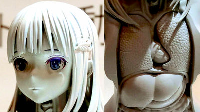
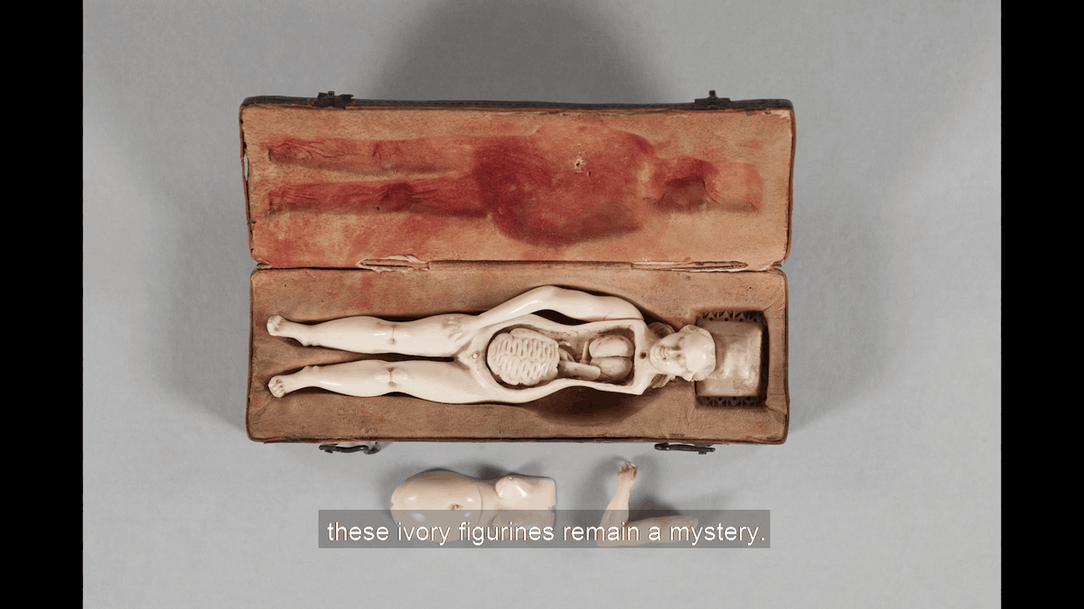
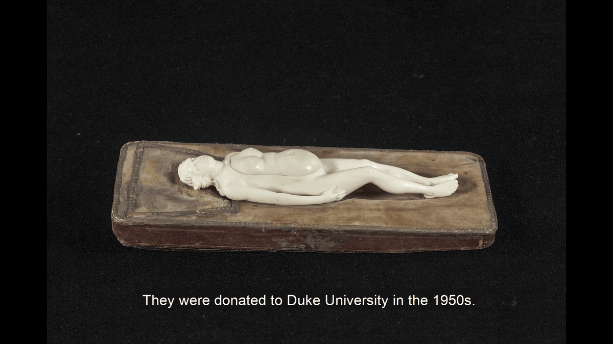
The 22 human models investigated this time were donated in 1956 from a personal collection of surgeons at Duke University Hospital. Each human body model is 12 to 24 centimeters, and depending on the thing, the arm seems to move.
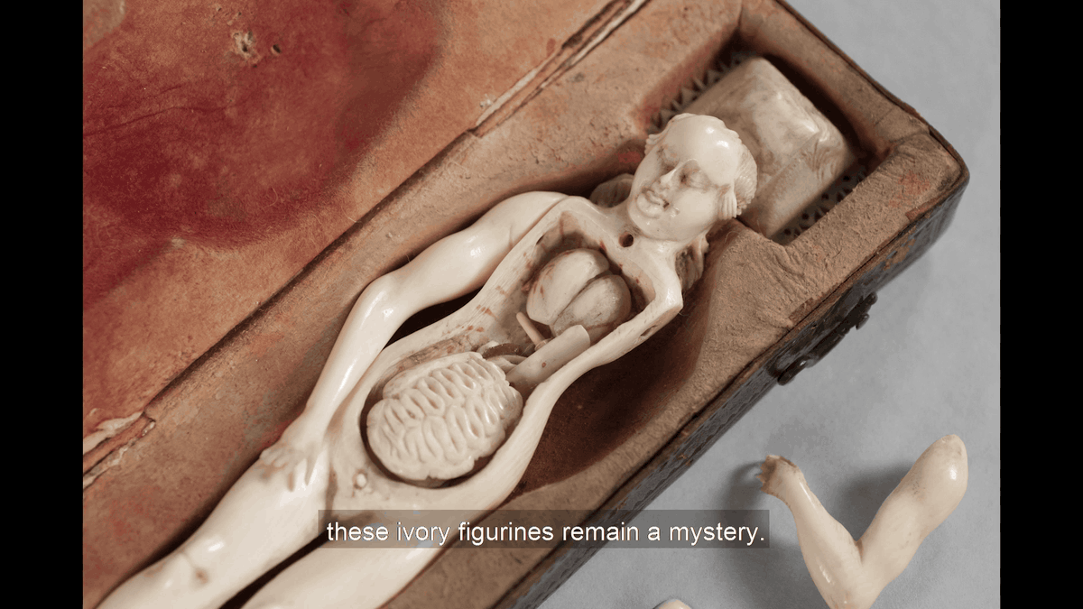
The body contains parts that reproduce the internal organs of the lungs and intestines.
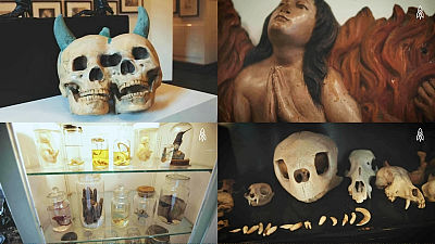
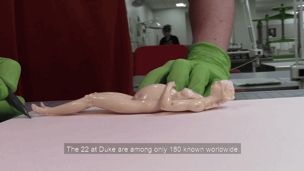
The following is another human model made at the same time. It is confirmed that there are 180 human models around the world, including 22 used in this survey. Since it is a human body model hundreds of years ago, it seems that some small parts were damaged or lost.

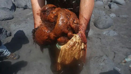
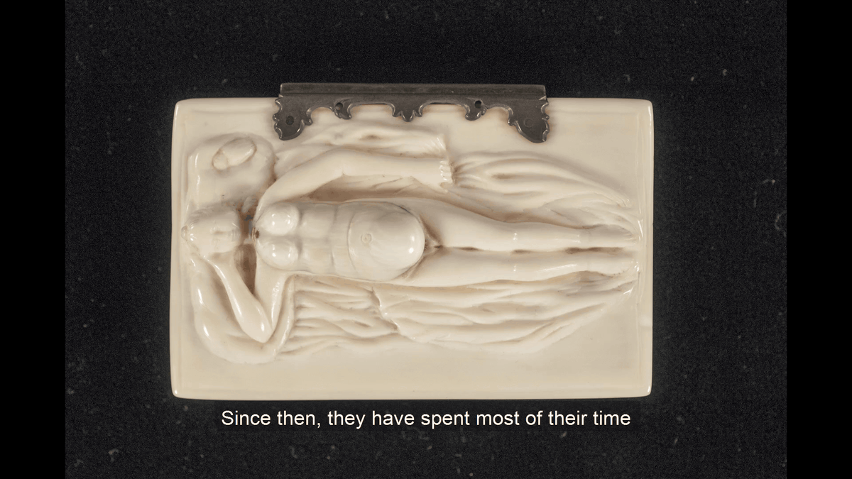
Female model with abdomen swelled.
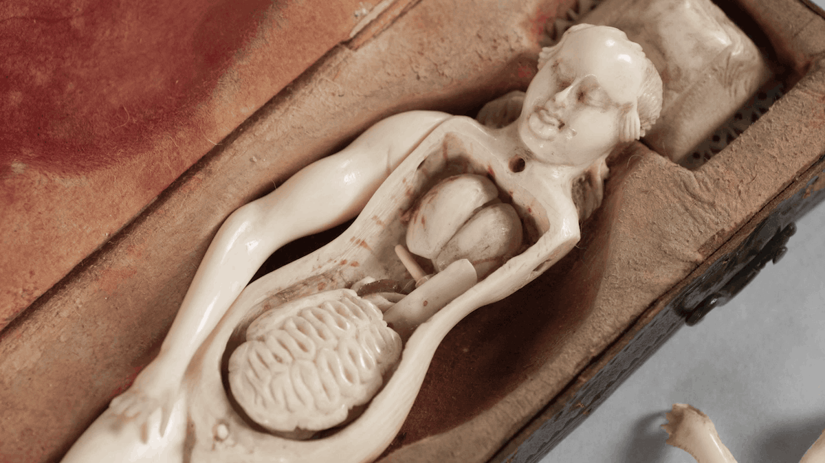
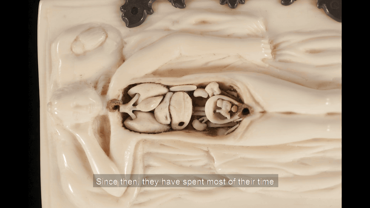
When the lid of the abdomen was opened, internal parts such as the diaphragm, stomach, kidney and uterus were housed. There is a fetus in the womb, and it is tied with a umbilical cord recreated with silk thread.

Some dug up a bed and a human body model from one ivory. Originally, the human body model is a three-dimensional object for medical use, but it has been pointed out that this human body model, which can be interpreted as “an image of a woman singing on a wrinkled sheet,” was an art object for viewing purposes. The
This human body model seems to have relatively little internal parts lost, and the abdomen was clogged with heart, lungs, bronchi, stomach, liver, kidneys, and uterus. The womb also contains small fetal parts.
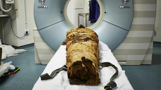
Researchers at Duke University trace the outline of the human body model for recording before performing a CT scan ...
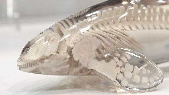
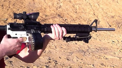
Scan with a micro CT scanner capable of CT scanning with higher resolution than usual.
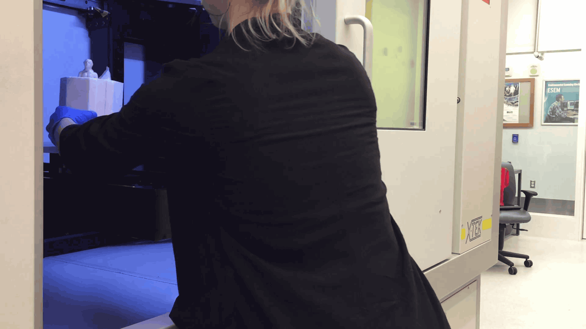
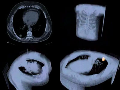
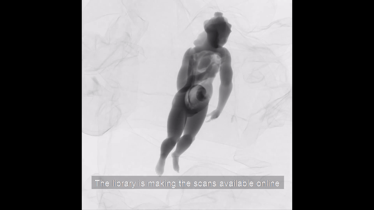
CT scans can be used to investigate the material and structure of the mannequin and to collect 3D data using X-rays. By taking X-rays from all angles, you can get detailed 3D data of the human body model.
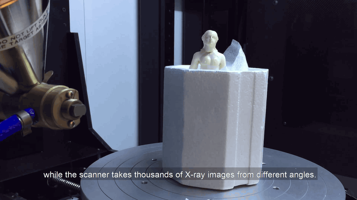
When actually shooting with X-rays, it looks like this. As a result of CT scan, 20 of the 22 human body models were made by carving ivory, one of the remaining 2 was made of deer bone, and the other was made of ivory and whale bone I understood that. In addition, it seems that there was a repaired trace depending on the human body model. The research team pointed out that ivory was a very difficult material to obtain in the second half of the 17th century when the human body model was made. This human model is said to be a clue to know what route ivory entered Europe from the 17th to 18th centuries.
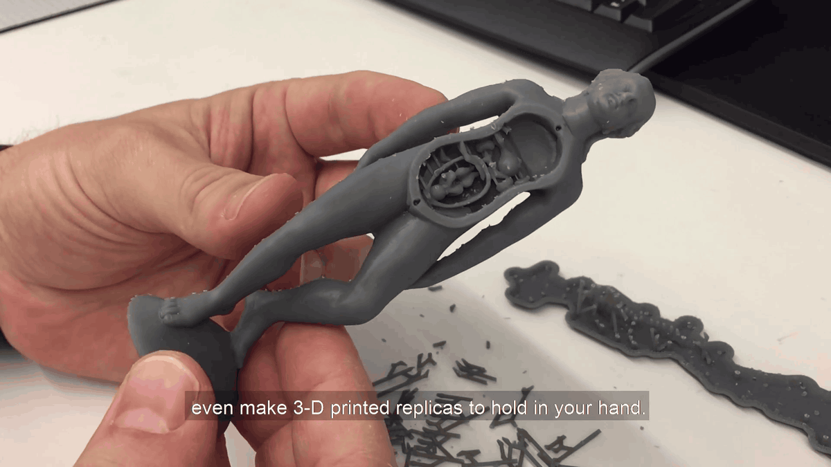
However, since all human models are very old and easily damaged, they are rarely exhibited at the Duke University medical library where they are stored. Therefore, the purpose of this survey is to save the shape of the human body model in digital format and output it with a 3D printer to create a replica for exhibition. Looking at the replica actually output by the 3D printer, it was finished with an accuracy that clearly shows the expression of the human body model and the fetus in the womb.
Related Posts: