If you shoot with the MRI scanner that has only three in the world how nerve signals are flowing in the brain
Brain scan images taken with a special MRI scanner that has only three in the world are released on BBC. This MRIAxonIt is possible to visualize how the neural signal is sent, it is possible to see in which direction the signal flows in a colorful image and how the density of the axon changes It is getting.
What the brain's squirrel looks like - BBC News
http://www.bbc.com/news/health-40488545
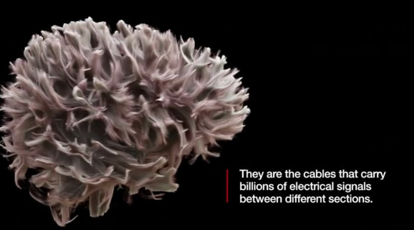
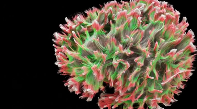
The following is an image taken with MRI scanner at Brain Research Imaging Center (CUBRIC) in Cardiff University, England. Of the brain, nerve fibers accumulateWhite matterRespectively. White matter tells billions of nerve signals from one part of the brain to another part.
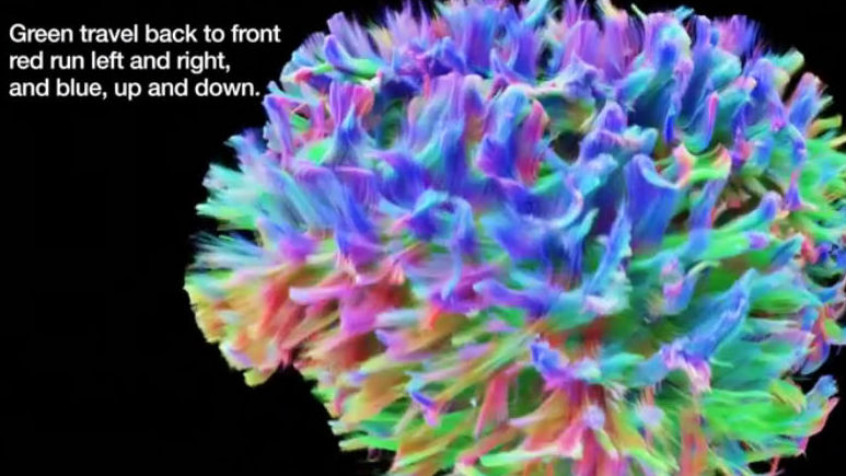
Various colors such as blue, red, green and so on represent the movement of the nerve signal. It seems that green indicates forward and backward movements, red indicates left and right movements, and blue indicates up and down movements.
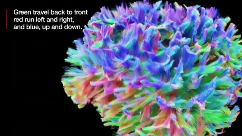
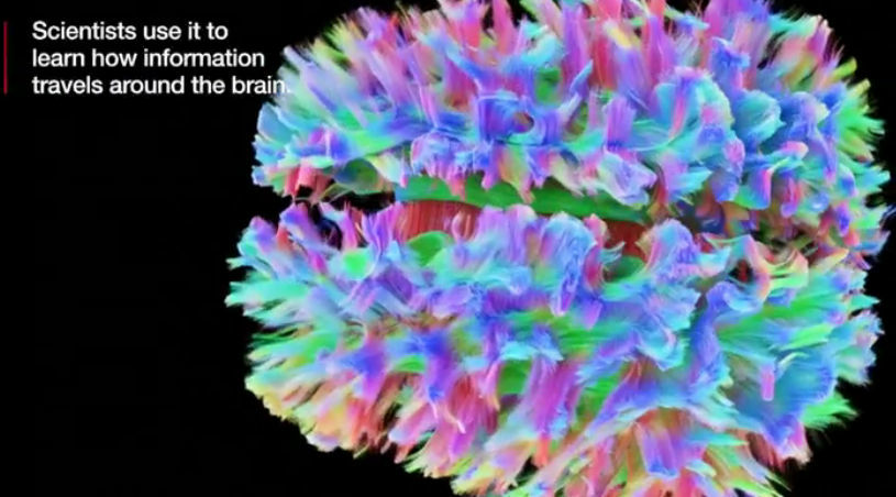
Looking at the brain from above it is like this. Especially, you can see that there are many parts of blue. With such coloring, researchers can learn how information is coming and going through the brain.
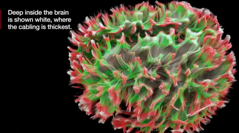
On the other hand, there are MRI images separated by two colors, red and green. Red is the part where the density of the nerve fiber decreases toward the outside of the brain, and green is the part with the medium density.
Deep inside the brain, where the nerves are dense, it is shown in white.
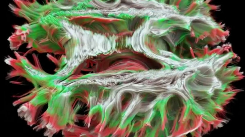
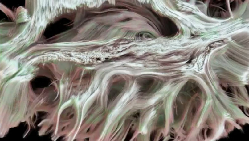
Close-up and close-out are also possible, as you get closer and closer ... ...
You can see how nerve fibers look like this. This technique is related to dementia and epilepsy,Multiple sclerosisIt is said to be used for investigation.
This MRI exam was taken by journalist Fergus Walsh. To Mr. Walsh, a medical correspondent at BBC, it was not special to take an MRI examination, and even when I was conducting an examination at CUBRIC, I could not find anything special to mention for 45 minutes. However, the images shown were amazing, showing up not only the direction of information transmission but also the density of brain connections. In MRI photography so far, even when photographing the brain of a person suffering from multiple sclerosis, only the place with functional impairment was understood. However, if you have the latest MRI scanner that CUBRIC has, the density of the axon is shown and you can see how the affected part affects the communication of information to the place where exercise and cognition is governed.
Professor Derek Jones of CUBRIC said that if the usual MRI is binoculars, this latest MRI is like a Hubble telescope and researchers will be able to see both structure and function for the first time at the first time is.
Related Posts:


in Science, Posted by darkhorse_log