A technology that improves the resolution of MRI by 64 million times compared to the past has appeared
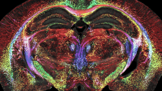
Magnetic resonance imaging (MRI), which is used to visualize soft tissue with a lot of moisture, which is difficult to capture with X-rays, has a voxel (minimum unit of a cube) of only 5 microns, which is 1/64 millionth of the previous level. A technique has been developed to express
Brain Images Just Got 64 Million Times Sharper | Duke Today
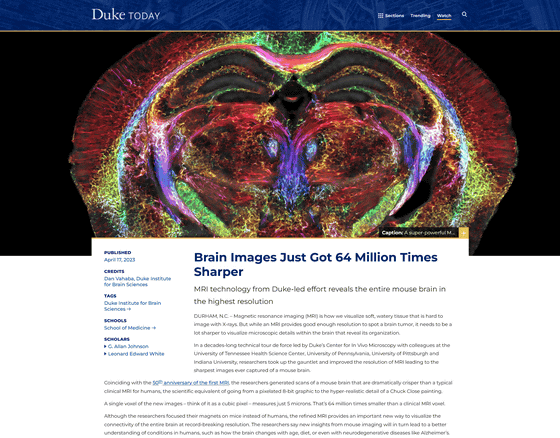
The technology is led by Duke University and created by teams from the University of Tennessee Health Science Center, the University of Pennsylvania, the University of Pittsburgh, and the University of Indiana.
MRI is used to visualize moist, soft tissue, such as the brain, and is useful for things like finding brain tumors, but it is still not sharp enough to visualize the details inside the brain. did not. The improved MRI has only 5 microns per voxel, which is 64 million times less than a conventional clinical MRI.
According to G. Alan Johnson, Ph.D., who has worked on the problem for more than 40 years at Duke University, the higher resolution is a combination of factors, including most clinical MRIs at 1.5
There are videos out there that show the difference.
Ultra-Sharp Brain Scan-YouTube
The left side of the black-and-white image is the conventional MRI, and the right side is the modified MRI. The details are completely different.

After scanning the brain, the tissue is imaged using a technique called
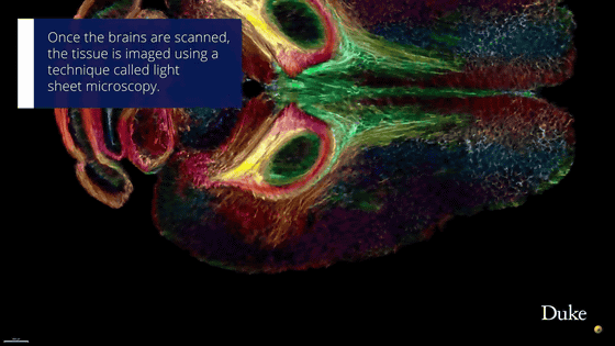
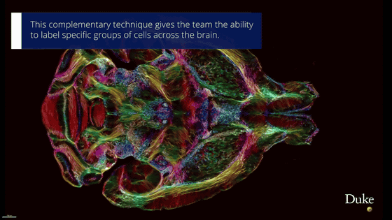
The light sheet image is then mapped onto the original MRI scan. This allows you to see anatomically accurate cells and circuits in your brain.
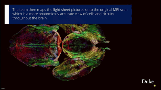
With this complementary technology, the research team is now able to label specific cell groups in the brain.
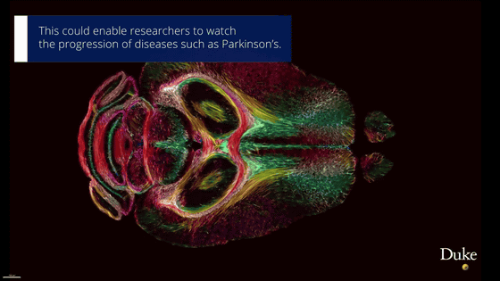
This makes it possible to monitor the progression of diseases such as Parkinson's disease.
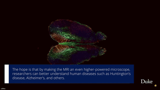
As MRI becomes a more powerful microscope, it is expected that we will be able to understand diseases such as Huntington's disease and Alzheimer's disease more deeply.
Related Posts:

