The first human-made 'full color 3D radiograph' realized with CERN technology

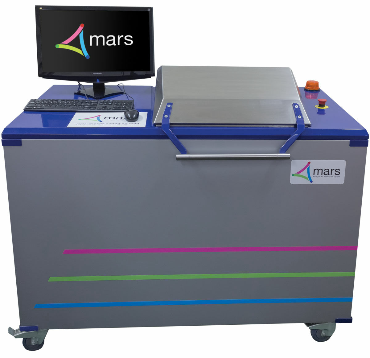
X-ray photography has become commonplace still in black and white still 100 years after discovery, but medical scanner " MARS " which can reproduce human body inside full color 3D model by shooting with X-ray Has been developed. For this latest X-ray scanner, the technology developed by the European Nuclear Research Institute (CERN) is applied.
First 3D color X-ray of a human using CERN technology | CERN
https://home.cern/about/updates/2018/07/first-3d-colour-x-ray-human-using-cern-technology
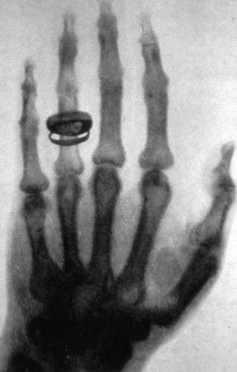
With the X-rays discovered by Wilhelm Röntgen in 1895, it became possible to show on the film what the living human beings are doing and to check them as pictures and images. Even in modern times more than 100 years since discovery, X-rays are used for healthcare sites and airport baggage inspections. Pictures and videos shot with X-rays can only be displayed in black and white in principle.
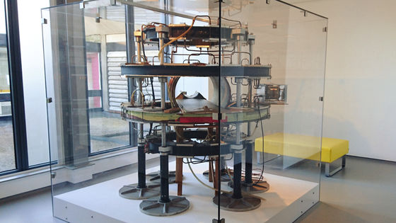
MARS Bioimaging Ltd. , which develops and sells 3D scanners, has developed a medical 3D scanner "MARS", which makes it possible to display X-ray photographs and images as full-color 3D models.
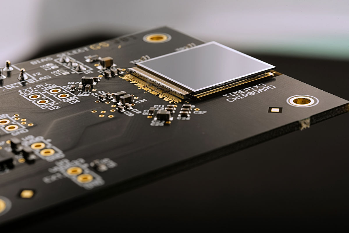
The chip set "Medipix" applied to this MARS was originally developed to trace particles with CERN's large hasron collision type accelerator, it can accurately detect particles colliding with the image sensor I will. Application of Medipix to the medical field was expected, because it makes it possible to shoot high resolution and high contrast X-ray photographs. MARS Bioimaging Ltd., in cooperation with various universities and research institutes, developed the third generation "Medipix 3" and applied it to MARS.
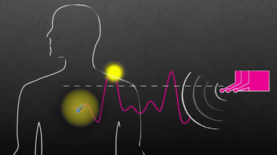
MARS analyzes spectral information of X-rays detected by Medipix 3 and generates full color 3D image. The X-ray attenuates the energy level when passing through a substance, but it discriminates fat, water, calcium, lesion part, etc. from this attenuated energy amount and reflects it on the color.
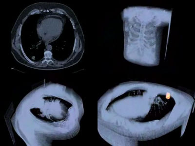
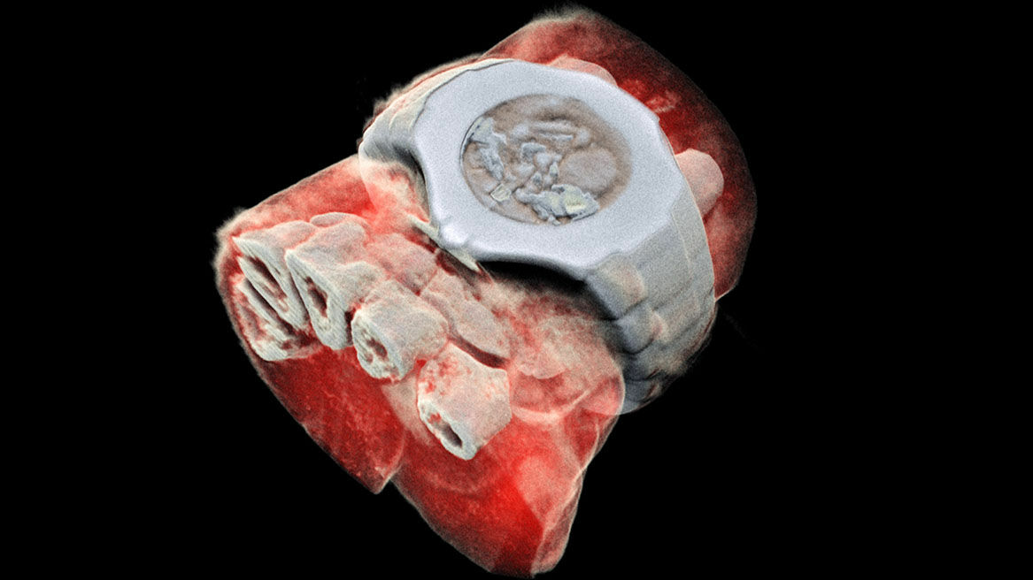
For example, in the following image, I scanned wrist wearing wristwatch with MARS. You can see that the bones of the wrist part are reproduced up to the cross section and are reflected.
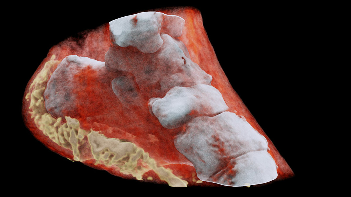
Also, looking at the image scanned by the MARS heel, you can see that there are yellow fats from the heel to the soles of the feet, besides the complex bones of the white foot bones.
According to CERN, a small version of MARS has also been developed, and it is being tested whether small version MARS can be used for diagnosis leading to early detection of bone and joint health status, cancer, and vascular disease at present And that. The development team considers the test results "promising". In addition, it is scheduled to be examined using a MARS scanner for patients with rheumatism as a clinical trial in Orthopedic Surgery in New Zealand. Aurelie Pezous, CERN's knowledge transfer, says, "It is a pleasure for us that CERN's research has benefited the world, and that the results of our research are applied to real life in this way is our CERN's I am encouraged by the research. "
Related Posts: