You may be able to easily output models of your organs with 3D printers
byRudi Aldridge
The doctor uses two-dimensional data such as scan images of MRI and CT to inspect the patient's symptoms. However, if it is possible to output a three-dimensional model which is three-dimensional data, not only doctors will help judge the symptoms, but also the patients themselves will greatly help them to accurately understand the condition of their bodies. Researchers at MIT and Harvard University are developing a new method to make 3D models quickly from MRI and CT scan data.
From Improved Diagnostics to Presurgical Planning: High-Resolution Functionally Graded Multimaterial 3D Printing of Biomedical Tomographic Data Sets | 3D Printing and Additive Manufacturing
https://www.liebertpub.com/doi/10.1089/3dp.2017.0140
Now, You Can Hold a Copy of Your Brain in the Palm of Your Hand - Neuroscience News
https://neurosciencenews.com/3d-brain-printing-9177/
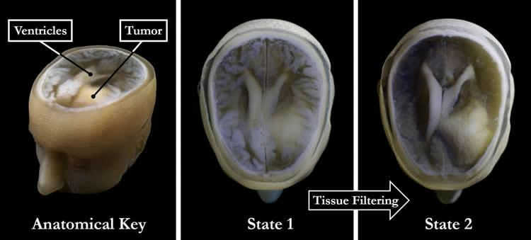
Steven Keatinging, who attended the graduate school of MIT, received a ball baseball tumor in the brain and underwent surgery to remove it. At this time, Keating, who felt wanting to know more about what is going on in his / her brain, collect scanned images of MRI or CT that photographed his / her brain and output it stereoscopically using a 3D printer He said that he tried. However, scanned images of MRI and CT show cross-sectional views obtained by cutting subjects into multiple layers, images are taken extremely precisely so that doctors and others can accurately determine the condition of the patient, and from the scan data 3D models , It is necessary to create a physical model for each layer, and it seems that enormous calculation processing was indispensable for 3D printing. Therefore, Mr. Keating started developing technology which can handle scan data as digital data more easily.
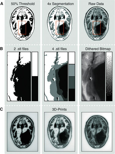
MIT Media Lab including Keating and the research group of Harvard University Wyss Institute have developed a new method of displaying scanned images in black and white bitmaps. In this method, as in black and white newspaper using black ink dots of different sizes in order to express shades, they made it possible to express shades by separately using black and white bitmaps. For example, by converting scanned images of MRI or CT with digital data such as bitmap, the shadow becomes darker if the number of black bitmaps is increased, it has been changed to a form that makes it easy to handle in 3D printers.
According to the method developed by researchers, we were able to produce solid models from MRI and CT scan data with overwhelming speed, but that is not the only merit. In scanned images of MRI and CT, processing that is called "segmentation" is performed in the body by separating the specific tissue from the surroundings so that the doctor can easily understand it. It is very time consuming for experts to perform segmentation manually. In some cases, segmentation is automatically performed by the computer, but since the irregular part is reflected in the actual scanned image or the boundary is not clear because the part including the gray area of the scanned image is reflected This work was difficult.
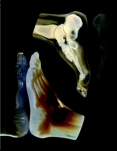
Using the method developed by researchers, we can handle automatic segmentation with overwhelmingly fast speed. "Even trained experts may take more than 30 hours to divide CT scanned images of human feet into bones, bone marrow, tendons, muscles, skin, etc. in the body, but in our approach So we can complete this task in less than an hour, "said Dr. James Weaver of the Wyss Institute.
Researchers believe that the method developed this time can be applied to the development of medical tools that make it easier for patients to understand their diagnosis results and make it easier to understand. Dr. Weaver says, "I think that patients who scan CT or MRI within 5 years will be able to acquire 3D models of their bodies in a few days."
Related Posts: